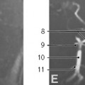
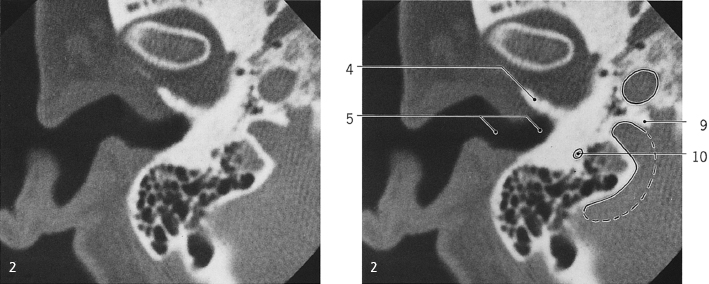
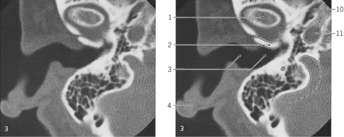
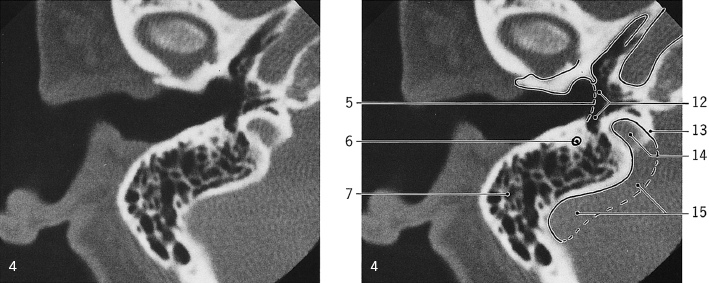
Petrous bone, CT series, diagrammatic scout view
Lines #1–14 indicate positions of sections in the following CT series. Consecutive sections, 3 mm thick
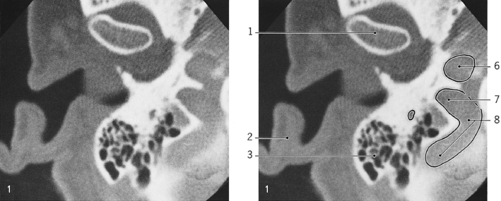
- Anterior semicircular canal
- Fenestra vestibuli
- Cochlea
- Auditory tube
- Carotid canal
- Mastoid antrum
- Posterior semicircular canal
- Lateral semicircular canal
- Pyramidal process
- Fenestra cochleae
- Facial canal
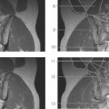
Ear, axial CT
Positions of sections are indicated above
- Head of mandible
- Auricle
- Mastoid process with air cells
- Tympanic part of temporal bone
- External acoustic meatus
- Carotid canal
- Bulb of internal jugular vein
- Sigmoid sinus
- Intrajugular process
- Facial canal
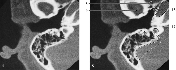
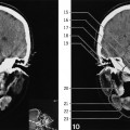
Ear, axial CT
Only gold members can continue reading.
Log In or
Register to continue

Stay updated, free articles. Join our Telegram channel