Innominate line (radiology term) (tangential view of greater wing of sphenoid bone)
Superior orbital fissure
Ethmoidal air cells
Maxillary sinus
Inferior nasal concha
Nasal septum
Dens axis
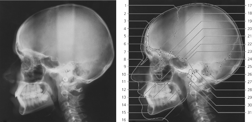
Skull, lateral X-ray
Coronal suture
Frontal bone
Outer table of calvaria
Diploë
Inner table of calvaria
Frontal sinus
Cribriform plate
Nasal bone
Ethmoidal air cells
Zygomatic process of maxilla
Maxillary sinus
Anterior nasal spine
Hard palate
Uvula
Mental protuberance
Angle of mandible
Parietal bone
Orbital plates of frontal bone
Greater wings of sphenoid bone
Jugum sphenoidale
Hypophysial fossa
Dorsum sellae
Sphenoidal sinus
Lambdoid suture
Occipitomastoid suture
Squamous part of occipital bone
Mastoid air cells
External acoustic meatus
Clivus
Mandibular neck
Anterior arch of atlas
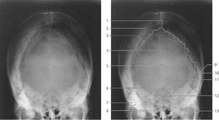
Skull, X-ray, Towne’s projection
Sagittal suture
Parietal bone
Lambdoid suture
Squamous part of occipital bone
Pineal gland (calcified)
Petrous part of temporal bone
Mastoid process
Nasal septum
Squamosal suture
Occipitomastoid suture
Foramen magnum
Sphenoidal sinus
Mandibular neck
Skull, lateral X-ray, old age
Granular foveolae
Grooves for branches of middle meningeal artery
Diploic veins
Pineal gland (calcified)
Lambdoid suture
Internal occipital protuberance
Air cells in temporal bone
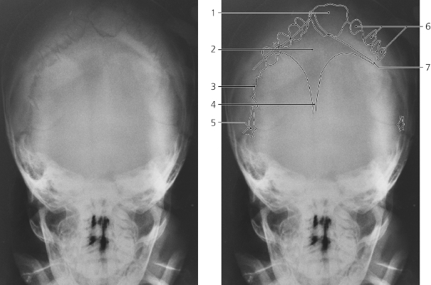
Skull, a-p, tilted X-ray, child 5 months
Interparietal bone (Inca bone)
Anterior fontanelle
Lambdoid suture
Sagittal suture
Mastoid fontanelle
Sutural (Wormian) bones in lambdoid suture
Coronal suture
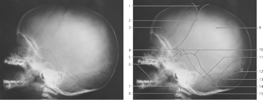
Skull, lateral X-ray, child 5 months
Anterior fontanelle
Coronal suture
Frontal bone
Pterion (sphenoidal fontanelle)
Greater wing of sphenoid bone
Deciduous teeth
Anterior arch of atlas
Dens axis
Parietal bone
Squamosal sutures
Mastoid fontanelles
Lambdoid suture
Sutural bone
Occipitomastoid suture
Posterior arch of atlas
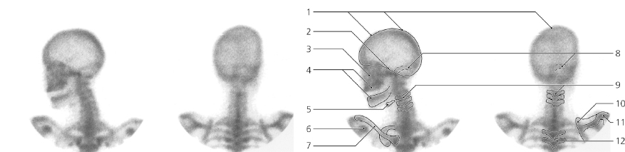
Skull, lateral and posterior view, 99m Tc-MDP, scintigraphy
Calvaria
Base of skull
Facial skeleton
Alveolar process of maxilla and alveolar part of mandible
Hyoid bone
Coracoid process
Clavicle
Transverse and sigmoid sinus
Cervical vertebra
Superior angle of scapula
Acromion
Thoracic vertebra
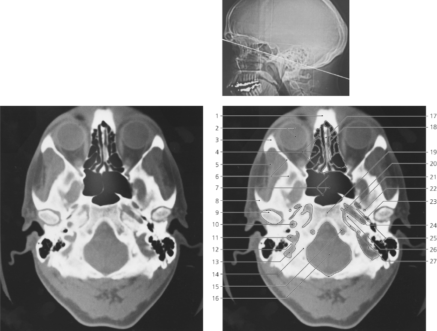
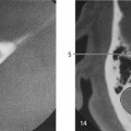
Base of skull, axial CT
Nasal spine of frontal bone
Eyeball
Frontal process of zygomatic bone
Ethmoidal air cells
Temporal fossa
Greater wing of sphenoid bone
Sphenoidal sinus
Zygomatic process of temporal bone
Head of mandible
Carotid canal, first part
Jugular foramen, posterior to intrajugular process
Posterior border of jugular foramen
Sigmoid sinus
Lateral part of occipital bone
Hypoglossal canal
Foramen magnum
Nasal septum
Nasal cavity
Body of sphenoid bone
Foramen lacerum
Foramen ovale
Foramen spinosum
Sphenopetrous fissure/ Eustachian tube
Carotid canal, second part
Air cells in temporal bone
Apex of petrous bone
Petro-occipital fissure
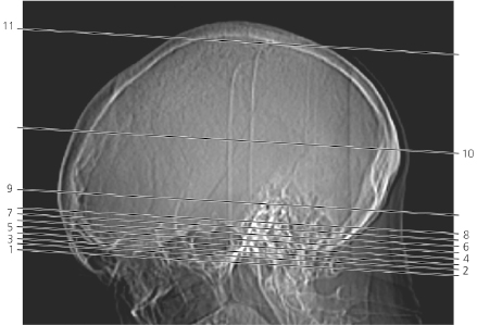
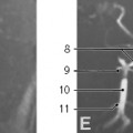
Scout view of skull
Lines #1–11 indicate position of sections in the following axial CT series displayed in bone settings. The corresponding series in brain settings is found on pages 245–7. This skull is highly pneumatized.
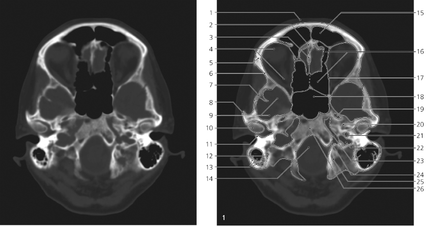
Skull, axial CT
Only gold members can continue reading. Log In or Register to continue