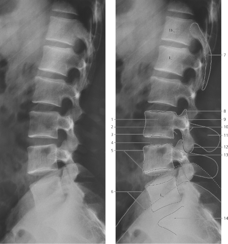
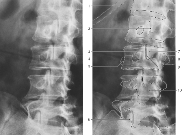
Lumbar spine, a-p X-ray
- Body of vertebra Th XII
- Head of 12th rib
- Spinous process of Th XII
- Upper and lower ambitus eminens of L I
- Superior articular process of L II
- Pedicle of vertebral arch L II
- Transverse process L II
- Lamina of vertebral arch L II
- Zygapophysial (facet) joint L II – L III
- Inferior articular process of L II
- Inferior articular process of L III
- Superior articular process of L IV
- Spinous process of L I
- Spinous process of L V
- Transverse process of L V
- Intervertebral disc L IV – L V
- Base of sacrum
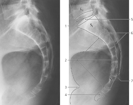
Lumbar spine, lateral X-ray
- Body of vertebra
- Intervertebral foramen
- Lower end plate of L III
- Intervertebral disc L III – L IV
- Upper and lower ambitus eminens
- Iliac crests
- 12th rib
- Superior articular process
- Pedicle of vertebral arch
- Lamina of vertebral arch
- Spinous process
- Inferior articular process
- Transverse (costal) process
- Sacrum
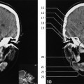
Lumbar spine, oblique X-ray
The “Scottie dog” projection
- 12th rib
- Zygapophysial (facet) joint L I – L II
- Superior articular process of L III
- Pedicle of vertebral arch L III (eye of “Scottie dog”)
- Transverse process of L III (snout of “Scottie dog”)
- Superior articular process of sacrum
- Inferior articular process of L II
- Transverse process of L III
- Zygapophysial (facet) joint L II – L III
- Lamina of vertebral arch L IV
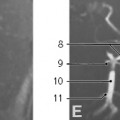
Sacrum, lateral X-ray
- Intervertebral disc L V – S I
- Pelvic surface of sacrum
- Ischial spine
- Coccyx
- Base of sacrum
- Sacral canal
- Sacral hiatus
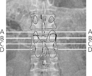
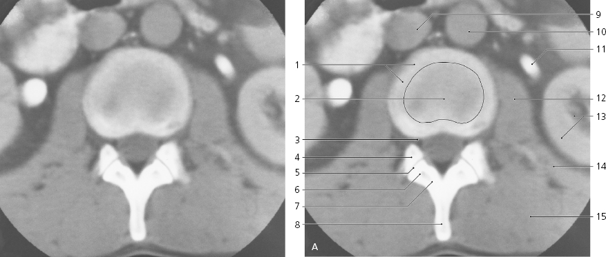
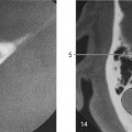
Lumbar spine, axial CT
Level of section A indicated on scout view above
- Anulus fibrosus of intervertebral disc
- Nucleus pulposus
- Intervertebral foramen for spinal nerve L II
- Superior articular process of L III
- Zygapophysial (facet) joint
- Inferior articular process of L II
- Lamina of vertebral arch
- Spinous process of L II
- Inferior caval vein
- Abdominal aorta
- Left ureter/pelvis (with contrast medium)
- Psoas major
- Left kidney
- Quadratus lumborum
- Erector spinae
Only gold members can continue reading.
Log In or
Register to continue

Stay updated, free articles. Join our Telegram channel