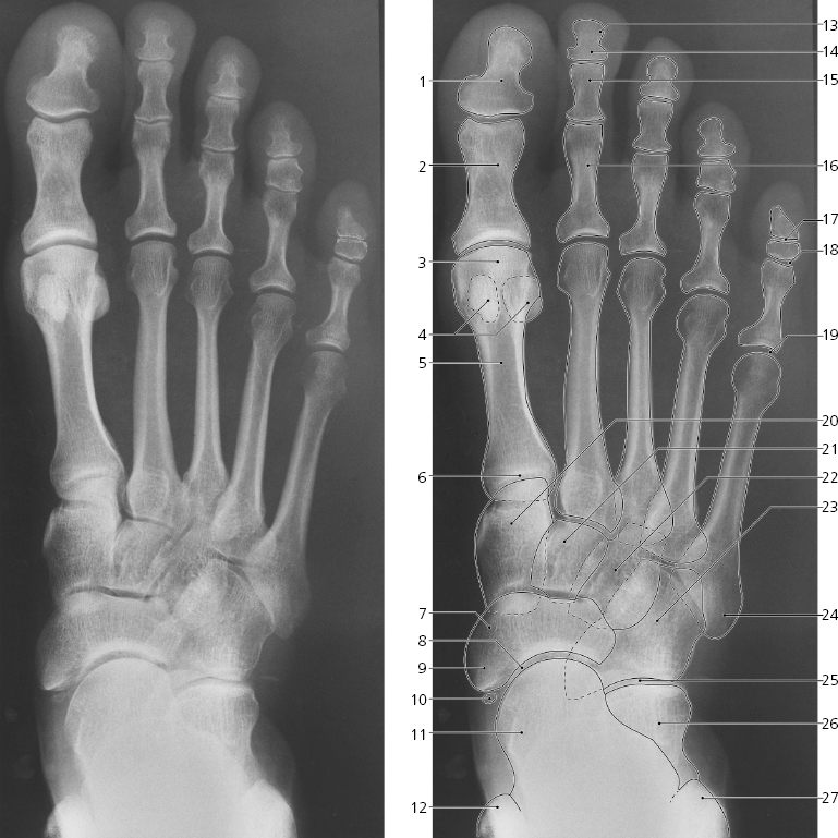
Sesamoid bone in tendon of flexor digitorum longus
Head of talus
Medial malleolus
Tuberosity of distal phalanx
Distal phalanx
Middle phalanx
Proximal phalanx
Distal interphalangeal joint (“DIP”)
Proximal interphalangeal joint (“PIP”)
Metatarsophalangeal joint (“MTP”)
Medial cuneiform bone
Intermediate cuneiform bone
Lateral cuneiform bone
Cuboid bone
Tuberosity of fifth metatarsal
Calcaneocuboideal joint
Calcaneus
Lateral malleolus
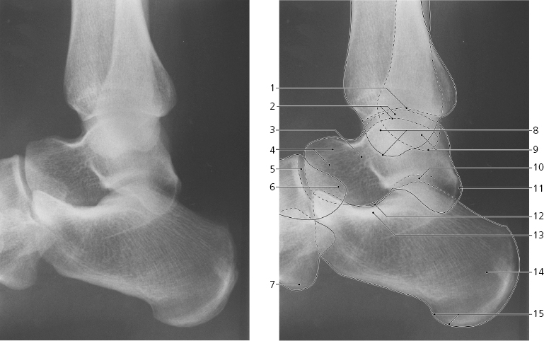
Foot, lateral X-ray
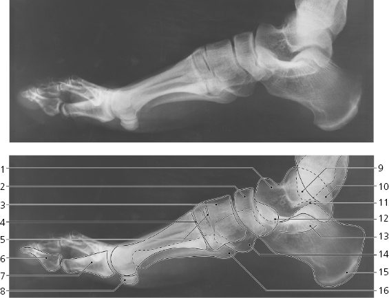
Head of talus
Navicular bone
Medial cuneiform bone
First tarsometatarseal joint
Second and third tarsometatarseal joints
Distal phalanx of great toe
Proximal phalanx of great toe
Sesamoid bones
Lateral malleolus
Medial malleolus
Subtalar joint
Tuberosity of navicular bone
Sustentaculum tali
Tuberosity of cuboid bone
Tuber calcanei
Tuberosity of fifth metatarseal
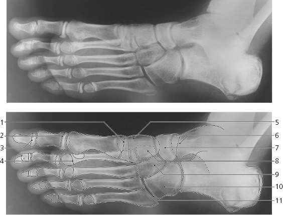
Foot, oblique X-ray
Growth plate of first metatarseal
Growth plate of proximal phalanx of great toe
Growth plate of distal phalanx of great toe
Growth plate of second metatarsal bone
Medial cuneiform bone
Head of talus
Navicular bone
Intermediate cuneiform bone
Lateral cuneiform bone
Cuboid bone
Fifth tarsometatarseal joint
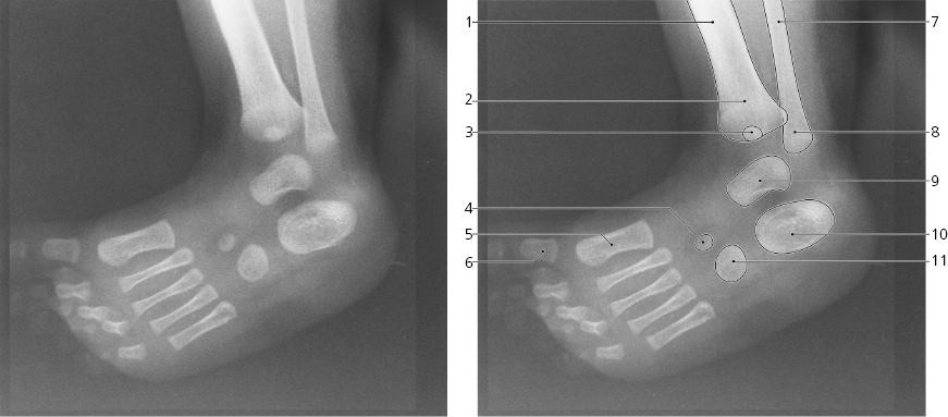
Foot, oblique X-ray, child 3 months
Diaphysis of tibia
Distal metaphysis of tibia
Distal epiphysis of tibia (ossification center)
Lateral cuneiform bone (ossification center)
Diaphysis of first metatarsal bone
Diaphysis of proximal phalanx of great toe
Diaphysis of fibula
Distal metaphysis of fibula
Talus (ossification center)
Calcaneus (ossification center)
Cuboid bone (ossification center)
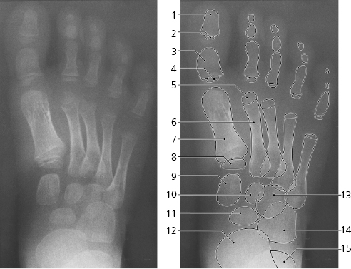
Foot, dorso-plantar X-ray, child 5 years
Diaphysis of distal phalanx
Epiphysis of distal phalanx
Diaphysis of proximal phalanx
Epiphysis of proximal phalanx
Epiphysis of second metatarsal bone
Diaphysis of second metatarsal bone
Diaphysis of first metatarsal bone
Epiphysis of first metatarsal bone
Medial cuneiforme bone
Intermediate cuneiforme bone
Navicular bone
Head of talus
Lateral cuneiforme bone
Cuboid bone
Calcaneus
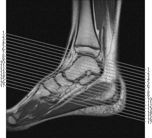
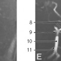
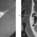
Scout view of ankle and foot
Lines #1–17 indicate position of sections in the following axial MR series. Interpretation of the scout image can be found in the sagittal series, page 167, image #8.
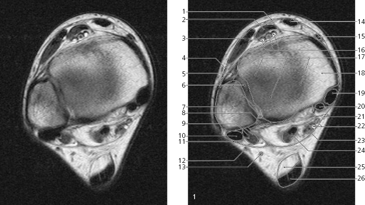
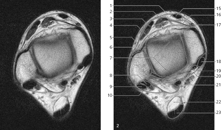
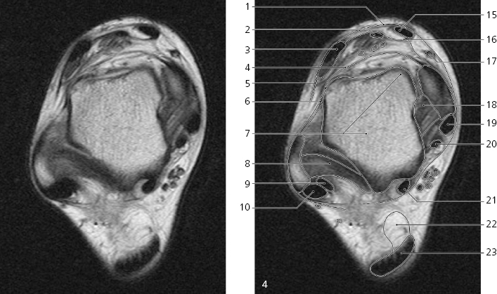
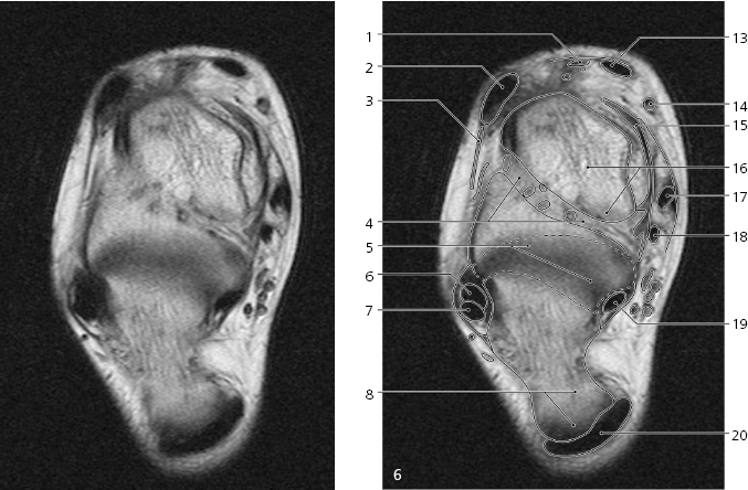
Ankle and foot, axial MR
Superior extensor retinaculum/ fascia cruris →
Extensor hallucis longus →
Extensor digitorum longus →
Peroneus tertius →
Anterior tibiofibular ligament →
Syndesmosis →
Lateral malleolus →
Posterior tibiofibular ligament →
Peroneus brevis →
Peroneus longus →
Superior peroneal retinaculum →
Small saphenous vein →
Sural nerve →
Tibialis anterior →
Great saphenous vein →
Dorsalis pedis artery and veins →
Articular cartilage of talocrural joint
Medial malleolus →
Tibialis posterior →
Flexor digitorum longus →
Flexor retinaculum →
Posterior tibial artery and vein →
Tibial nerve →
Flexor hallucis longus →
Karger’s fat pad →
Calcaneal tendon (Achilles) →
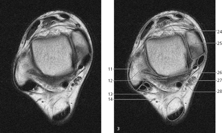
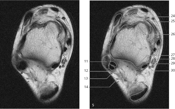
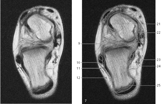
Ankle and foot, axial MR
Scout view on page 150
Superior extensor retinaculum/ fascia cruris ↔
Extensor hallucis longus ↔
Extensor digitorum longus ↔
Anterior articular capsule →
Peroneus tertius ↔
Anterior tibiofibular ligament (lower edge) ←
Trochlea tali →
Peroneus brevis (muscle and tendon) ↔
Peroneus longus ↔
Superior peroneal retinaculum ↔
Lateral malleolus ↔
Posterior articular capsule and syndesmosis tibiofibulare