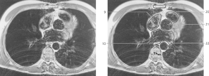
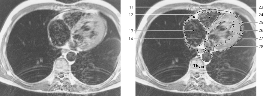
Heart, axial MR, level Th VI, Th VII and Th VIII
T1 weighted recording
- Body of sternum
- Internal thoracic artery and vein
- Ascending aorta
- Superior caval vein
- Left atrium
- Esophagus
- Azygos vein
- Thoracic duct
- Right atrium
- Right inferior pulmonary vein
- Right ventricle
- Right coronary artery
- Right atrium
- Interatrial septum
- Anterior mediastinum (sternopericardial ligament)
- Pulmonary trunk
- Left auricle
- Root of left lung
- Thoracic aorta
- Conus arteriosus
- Bulb of aorta
- Left inferior pulmonary vein
- Interventricular septum
- Left ventricle
- Pericardial sac
- Pericardial cavity
- Myocardium of left ventricle
- Left atrium
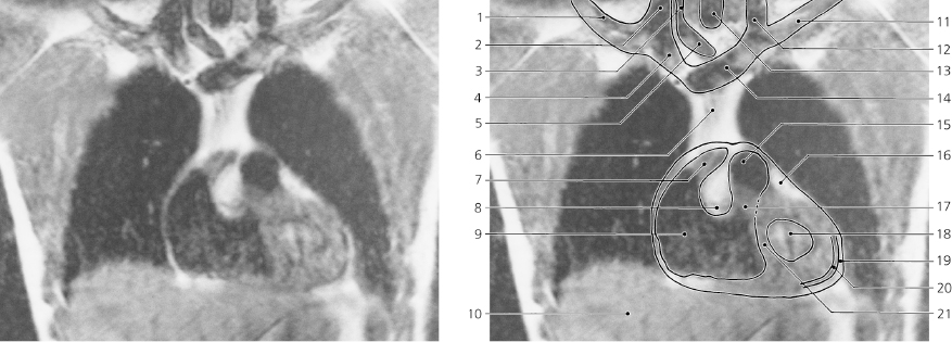
Heart, coronal MR
T1 weighted recording
- Right subclavian vein
- Right internal jugular vein
- Right common carotid artery
- Right brachiocephalic vein
- Brachiocephalic trunk
- Superior mediastinum with thymus
- Right atrium
- Supraventricular crest
- Right ventricle
- Liver
- Left subclavian vein
- Left internal jugular vein
- Trachea
- Left brachiocephalic vein
- Pulmonary trunk
- Epicardial fat
- Conus arteriosus
- Left ventricular cavity
- Pericardial sac
- Pericardial cavity
- Interventricular septum
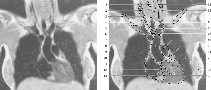
Heart, coronal MR
T1 weighted recording
- Body of cervical vertebra
- Right internal jugular vein
- Right common carotid artery
- Clavicle
- Right subclavian vein
- Right brachiocephalic vein
- Superior caval vein
- Ascending aorta
- Aortic valve
- Right atrium
- Right atrial wall, pericardium and pleura
- Interventricular septum, membranous part
- Interventricular septum, muscular part
- Left common carotid artery
- Left internal jugular vein
- Trachea
- Left brachiocephalic vein
- Brachiocephalic trunk
- Pulmonary trunk
- Left auricle
- Left ventricle
- Myocardium of left ventricle
- Mamma
- Right ventricle
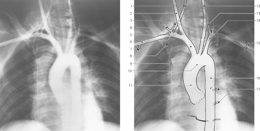
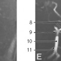
Aortic arch and great arteries, a-p X-ray (slightly oblique), aortography
- Right vertebral artery
- Right common carotid artery
- Inferior thyroid artery
- Transverse cervical artery
- Right subclavian artery
- Axillary artery
- Subscapular artery
- Internal thoracic artery
- Brachiocephalic trunk
- Aortic arch
- Ascending aorta
- Left common carotid artery
- Left vertebral artery
- Left subclavian artery
- Thoraco-acromial artery
- Thoracic aorta
- Intercostal arteries
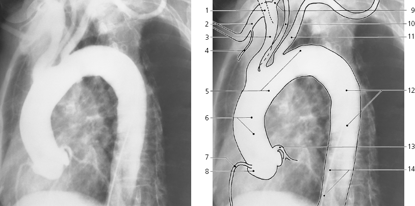
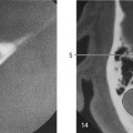
Aortic arch and great arteries, oblique X-ray, aortography
- Right common carotid artery
- Right subclavian artery
- Brachiocephalic trunk
- Internal thoracic artery
- Aortic arch
- Ascending aorta
- Right coronary artery
- Aortic sinus
- Right vertebral artery
- Left common carotid artery
- Left subclavian artery
- Thoracic aorta
- Left coronary artery
- Catheter
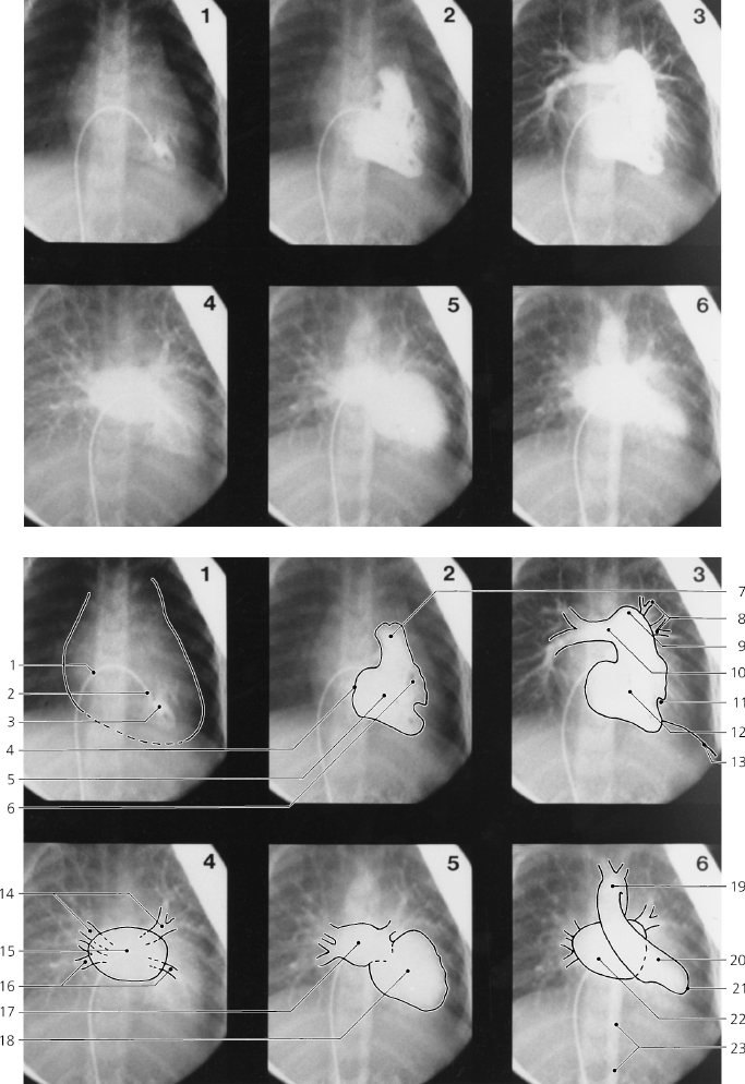
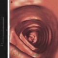
Heart, a-p, cardiac cineangiography, child
Only gold members can continue reading.
Log In or
Register to continue

Stay updated, free articles. Join our Telegram channel