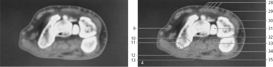
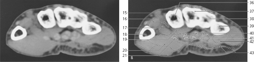
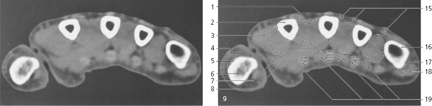
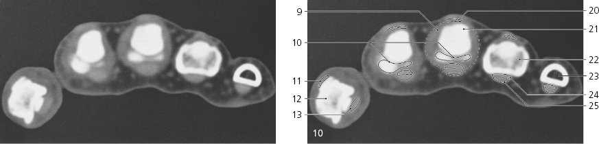
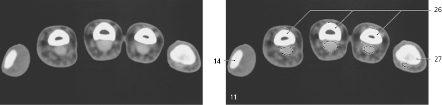
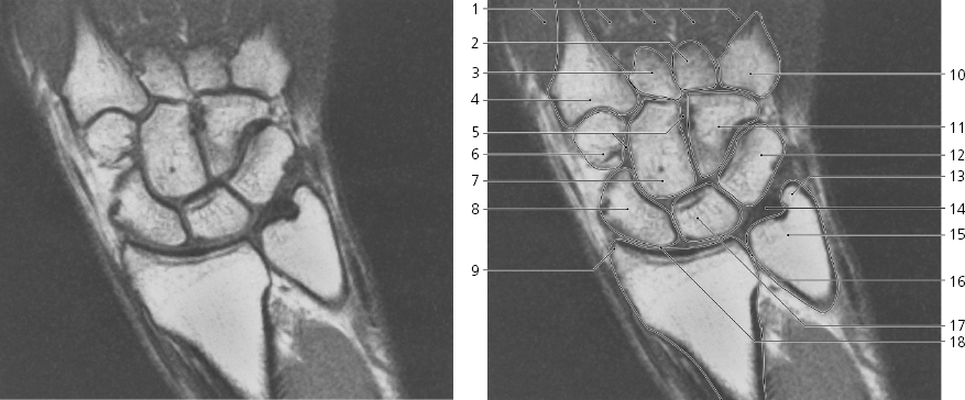
Lines # 1-11 indicate position of sections (1.5 mm thick) in the following CT series. Arrows ←, →, and ↔ in the legends indicate that a structure can be seen on a previous or following section, or both.
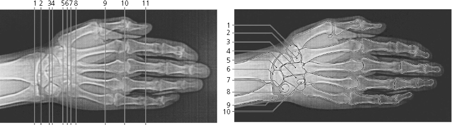
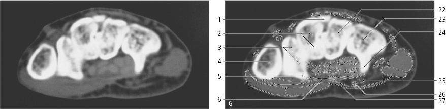
Trapezium
Trapezoid bone
Capitate bone
Hamate bone
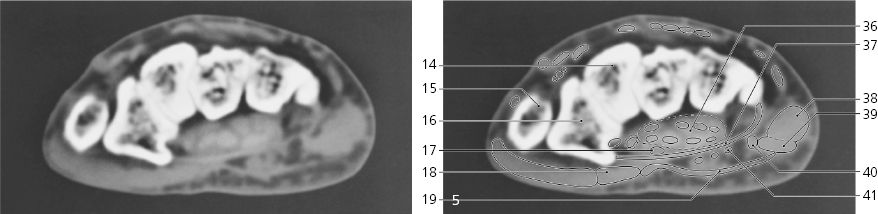
Scaphoid bone
Lunate bone
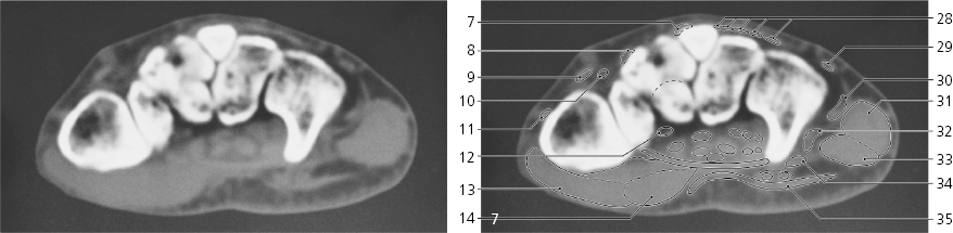
Pisiform bone
Triquetrum bone
Styloid process of ulna
Hook of hamate bone
Wrist, axial CT
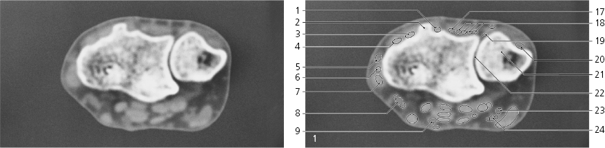
Extensor pollicis longus (tendon) →
Dorsal tubercle of radius
Extensor carpi radialis brevis (tendon) →
Extensor carpi radialis longus (tendon) →
Cephalic vein →
Extensor pollicis brevis (tendon) →
Abductor pollicis longus (tendon) →
Radial artery and veins →
Median nerve →
Distal edge of radius
Scaphoid bone →
Styloid process of radius
Joint capsule with palmar radiocarpal ligament
Flexor pollicis longus (tendon) ↔
Flexor carpi radialis (tendon) ↔
Palmaris longus (tendon) ↔
Extensor indicis (tendon) →
Extensor digitorum (tendons) →
Extensor digiti minimi (tendon) →
Extensor carpi ulnaris (tendon) →
Head of ulna
Distal radio-ulnar joint
Ulnar nerve →
Ulnar artery and veins →
Basilic vein ↔
Articular disc
Styloid process of ulna
Lunate bone →
Flexor digitorum profundus (tendons) ↔
Flexor carpi ulnaris (tendon) ↔
Flexor digitorum superficialis (tendons) ↔
Wrist, axial CT
Scout view on page 79
Extensor carpi radialis brevis (tendon) ↔
Extensor pollicis longus (tendon) ↔
Extensor carpi radialis longus (tendon) ↔
Articular capsule
Extensor pollicis brevis (tendon) ↔
Abductor pollicis longus (tendon) ↔
Radial artery and veins ↔
Abductor pollicis brevis →
Cephalic vein ↔
Flexor pollicis longus (tendon) ↔
Flexor carpi radialis (tendon) ↔
Tubercle of scaphoid bone
Palmaris longus (tendon) ←
Trapezoid bone →
Base of first metacarpal bone
Trapezium →
Median nerve ↔
Flexor pollicis brevis →
Palmar aponeurosis →
Extensor retinacle
Basilic vein ↔
Lunate bone ←
Triquetrum bone →
Capitate bone →
Scaphoid bone ↔
Pisiform bone →
Ulnar artery and veins ↔
Extensor indicis and digitorum (tendons) ↔
Extensor digiti minimi (tendon) ↔
Extensor carpi ulnaris (tendon) ↔
Hamate bone →
Flexor digitorum profundus (tendons) ↔
Flexor digitorum superficialis (tendons) ↔
Flexor carpi ulnaris (insertion) ←
Ulnar nerve ↔
Common synovial sheath of digital flexors ↔
Flexor retinacle ↔
Abductor digiti minimi →
Flexor digiti minimi →
Pisometacarpeal ligament →
Pisohamate ligament
Wrist, axial CT
Scout view on page 79
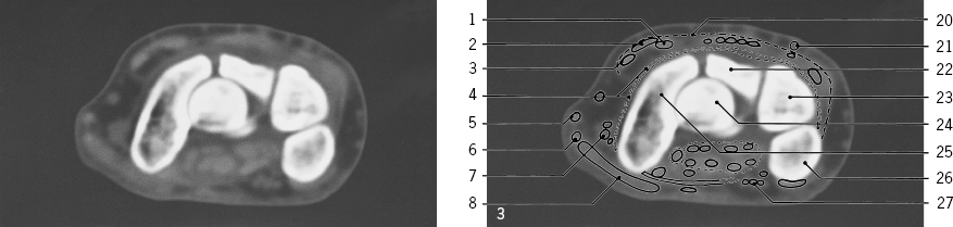
Styloid process of third metacarpal bone
Trapezoid bone ←
Trapezium ←
Base of first metacarpal bone ↔
Tubercle of trapezium
Flexor retinacle ↔
Extensor carpi radialis brevis (insertion) ←
Extensor carpi radialis longus (insertion) ←
Extensor pollicis longus (tendon) ↔
Radial artery ↔
Extensor pollicis brevis (tendon) ↔
Flexor carpi radialis (tendon) ←
Abductor pollicis brevis ↔
Flexor pollicis brevis ↔
Radial artery (turning into deep palmar arch) ←
First dorsal interosseus muscle →
Flexor pollicis brevis, deep head
Shaft of first metacarpeal bone ↔
Adductor pollicis →
Opponens pollicis ←
Flexor pollicis longus (tendon) ↔
Capitate bone ↔
Hamate bone ↔
Hook of hamate bone
Ulnar nerve ↔
Ulnar artery ↔
Median nerve ↔
Extensor indicis and digitorum (tendons) ↔
Extensor digiti minimi (tendon) ↔
Extensor carpi ulnaris (tendon) ←
Abductor digiti minimi ↔
Pisometacarpeal ligament ←
Flexor digiti minimi ↔
Palmar carpometacarpeal ligament
Palmar aponeurosis ↔
Base of second metacarpal bone →
Base of third metacarpal bone →
Base of fourth metacarpal bone
Base of fifth metacarpal bone
Flexor digitorum profundus (tendons) ↔
Flexor digitorum superficialis (tendons) ↔
Opponens digiti minimi ←
Palmaris brevis
Metacarpus and fingers, axial CT
Scout view on page 79
Second dorsal interosseus muscle ←
Shaft of second metacarpal bone ←
First dorsal interosseus muscle ←
Extensor pollicis longus (tendon) ↔
Extensor pollicis brevis (insertion) ←
Proximal phalanx of thumb
Adductor pollicis ←
First palmar interosseus muscle
Joint capsule of third carpometacarpeal joint
Fibrocartilaginous plates of palmar ligament
Extensor pollicis longus (insertion) ←
Distal phalanx of thumb
Flexor pollicis longus (tendon) ←
Tuberosity of distal phalanx
Veins
Head of fifth metacarpal bone
Flexor digiti minimi ←
Abductor digiti minimi ←
Lumbrical muscles
Extensor digitorum (tendon) ↔
Head of third metacarpal bone
Base of proximal phalanx of fourth finger
Shaft of proximal phalanx of fifth finger
Flexor digitorum profundus ↔
Flexor digitorum superficialis ↔
Shafts of proximal phalanges of second, third and fourth finger
Base of middle phalanx of fifth finger
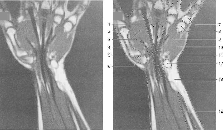
Wrist, coronal MR
Interossei muscles
Base of fourth metacarpal bone
Base of third metacarpal bone
Base of second metacarpal bone
Interosseous ligaments
Trapezoid bone
Capitate bone
Scaphoid bone
Styloid process of radius
Base of fifth metacarpal bone
Hamate bone
Triquetrum bone
Styloid process of ulna
Articular disc
Head of ulna
Distal radio-ulnar joint
Lunate bone
Radiocarpal joint
Wrist, carpal tunnel, coronal MR
Lumbricals
Head of first metacarpal bone
Flexor pollicis brevis, and adductor pollicis
Flexor pollicis longus (tendon)
Base of first metacarpal bone
Trapezium
Proximal phalanx of fifth finger
Head of fifth metacarpal bone
Flexor digiti minimi
Abductor digiti minimi
Long flexor tendons in canalis carpi
Pisiform bone
Subcutaneous fat
Shaft of ulna
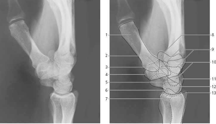
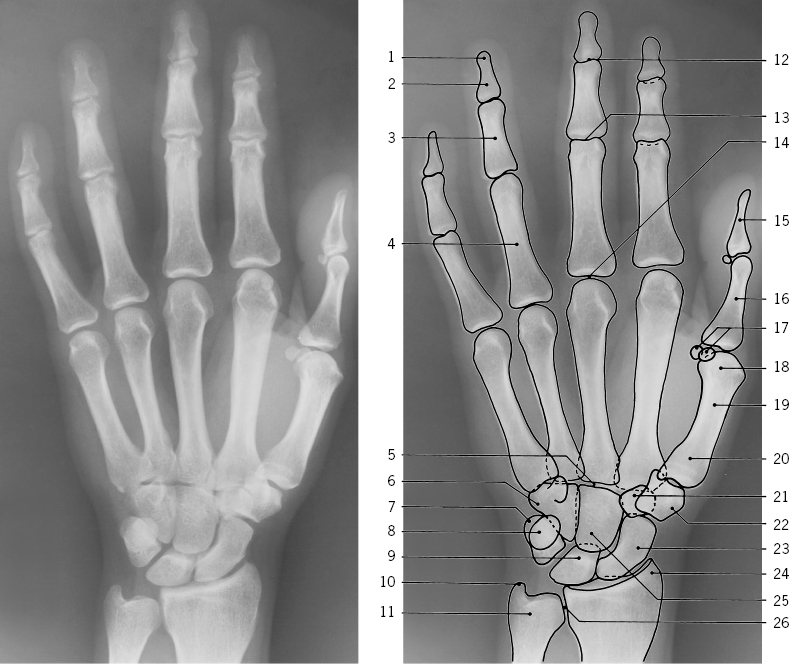
Hand, left, dorso-volar X-ray
Tuberosity of distal phalanx
Distal phalanx
Middle phalanx
Proximal phalanx
Carpometacarpeal joint
Hamate bone
Triquetrum bone
Pisiform bone
Lunate bone
Styloid process of ulna
Head of ulna
Distal interphalangeal joint “DIP”
Proximal interphalangeal joint “PIP”
Metacarpophalangeal joint “MCP”
Distal phalanx of thumb
Proximal phalanx of thumb
Sesamoid bones
Head of first metacarpal bone
Shaft of first metacarpal bone
Base of first metacarpal bone
Trapezoid bone
Trapezium
Scaphoid bone
Styloid process of radius
Capitate bone
Distal radio-ulnar joint
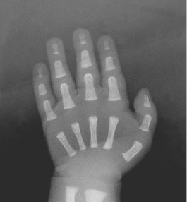
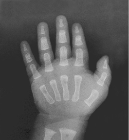
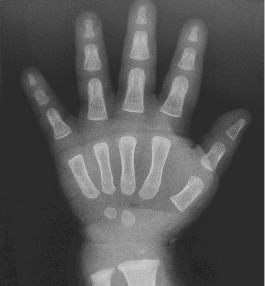
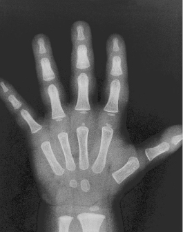
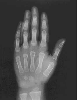
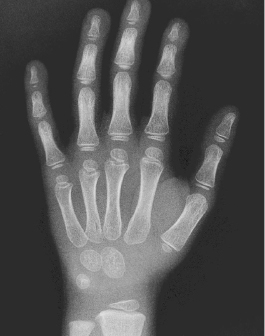
Skeletal age of hand The skeletal development of the hand of boys and girls is displayed on the following pages 85–92 The skeletal (bone) age of each hand (left) is given according to Greulich and Pyle (1) (upper line), and according to the 20 bone scoring system of Tanner et al. (2) followed by the 10 to 90 centile interval of variation (lower line).
W.W. Greulich and S.J. Pyle: Radiographic atlas of skeletal development of the hand and wrist. Stanford University Press 1959.
J.M. Tanner, R.H. Whitehouse, N. Cameron, W.A. Marshall, M.J.R. Healy and H. Goldstein: Assessment of skeletal maturity and prediction of adult height (TW2 method). Academic Press 1983.
Boy, newborn 0 yearsBoy, ½ yearBoy, 1 yearBoy, 1 ½ year year ()Boy, 2 years 2 years ()Boy, 3 years years (
 year (
 )
)
 )
)
 years (