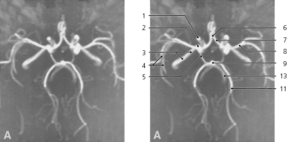
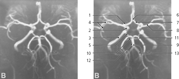
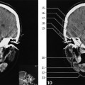
The following MR angiography series shows the bilateral set of cerebral arteries in a volume of brain, limited anteriorly and posteriorly, cranially and caudally as indicated by the frame on the scout view. The individual images A-E are the projected views perpendicular to the planes indicated by A-E on the scout view. See corresponding series on pages 318–319.
Brain arteries, MR angiography, circle of Willis
Internal carotid artery, “siphon”
Internal carotid artery in cavernous sinus
Internal carotid artery in carotid canal
Insular branches of middle cerebral artery
Posterior communicating artery
Anterior communicating artery
Anterior cerebral artery
Middle cerebral artery
Basilar artery
Superior cerebellar artery
Posterior cerebral artery
Anterior inferior cerebellar artery (AICA)
Vertebral artery
Only gold members can continue reading. Log In or Register to continue