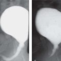
Anomalous Renal Size


10.1055/b-0034-87876

Anomalous Renal Size


Unilateral Small Kidneys


A kidney is small if it is > 2 standard deviations (SDs) below normal size for age. Causes may be congenital, secondary to abnormal development, or acquired due to parenchymal loss from a multitude of factors. The remaining normal renal tissue can undergo compensatory hypertrophy. Postnatally, this can be seen 12 to 18 months later ( Tables 3.1 and 3.2 ).


Table 3.1 Unilateral small kidneys without pelvicaliceal (PC) dilatation

Diagnosis


Findings


Comments


Congenital hypoplasia (rare)


US: normal echogenicity and corticomedullary differentiation.


Voiding cystourethrogram (VCUG): normal. Excretory urography (EU): often decreased number of calyces.


Nuclear medicine (NM): Decreased function. Scars suggest segmental hypoplasia/infection.


Small kidney, normal calyces, but often fewer in number. Normal histology.


No familial link Segmental hypoplasia (Ask-Upmark kidney): Can lead to severe hypertension. May actually be secondary to infection.


Dysplasia


Fig. 3.1, p. 240


Fig. 3.2, p. 240


Can involve all or part of a kidney and can be bilateral. US: Increased echogenicity, loss of corticomedullary differentiation. Multiple cortical cysts (small to large). Doppler: increased resistive index (RI) in affected segment.


EU: May show ectopic insertion of ureter. May have dilated PC system and ureter from vesicoureteral reflux (VUR) or obstruction.


NM: Non- or poorly functioning kidney depending on the extent of dysplasia.


Can be associated with ureteral agenesis or atresia; ureteropelvic junction obstruction; ectopic ureteric insertion (with distance from normal insertion proportional to dysplasia).


Can be secondary to obstruction in the first half of pregnancy.


Often associated with VUR.


Renal artery stenosis


Fig. 3.3, p. 240


Fig. 3.4, p. 240


US: Decrease in size of all or part of kidney.


Doppler: Increased velocity (> 180 cm/s) immediately distal to the stenosis. Tardus et parvus waveform on intrarenal arteries.


Decreased diastolic flow.


NM: Delayed perfusion and decreased maximal peak. Increased sensitivity with captopril. Angiography/MRI/CT: narrowing of renal artery.


Associated with fibromuscular hyperplasia, neurofibromatosis, radiation arteritis, and Takayasu disease. Multiple renal arteries may be present and not all affected.


Atrophy secondary to renal artery occlusion


Doppler: absent blood flow with capsular collaterals. Contrast-enhanced CT (CECT): Nonenhancement. Absent or small renal artery.


Causes: thrombotic/thromboembolic, umbilical artery catheter, postsurgery.


Atrophy secondary to radiation


Smooth renal outline with cortex and medulla affected.


Radiation > 30 Gy will result in renal damage.


Atrophy secondary to renal vein thrombosis


US/CT/MRI: Secondary calcification of small intrarenal veins (dystrophic calcification around pyramids). Collateral veins at renal hilum. Renal vein may recanalize.


Can be bilateral.

Fig. 3.1 MCDK. US image of a MCDK showing multiple cysts of varying sizes with no discernible intervening renal parenchyma.
Fig. 3.2 MCDK. Coronal T2-weighted image of the abdomen shows multiple cysts of varying sizes in the right kidney. A nonfunctioning kidney was noted on nuclear scan.
Fig. 3.3 Renal artery stenosis. MRI coronal image: smaller hypoperfused right kidney.
Fig. 3.4 Severe right renal artery stenosis is seen on digital subtraction angiography. Note the delay in the arterial phase of opacification between the two kidneys.


Table 3.2 Unilateral small kidney with PC dilatation

Diagnosis


Findings


Comments


Dysplastic kidneys


(see unilateral dysplastic kidneys in Table 3.1 )


Renal dysplasia associated with in-utero ureteric obstruction or reflux.


Postobstructive atrophy


US: Dilated PC system. Thinned cortex with increased echogenicity.


CT/MRI: can show level of obstruction.


Can be associated with dysplasia especially if prenatal obstruction.


Decreased size due to pyelonephritis


Fig. 3.5


US: small kidney, asymmetric scarring/cortical thinning over calyces.


NM (dimercaptosuccinic acid [DMSA]): focal scarring with photopenic areas and decreased function.


MRI: dynamic postcontrast imaging can show scarring as areas of cortical thinning and decreased contrast uptake. Can also assess PC system, ureters, split renal function, and glomerular filtration rate (GFR).


Also known as “reflux nephropathy.” VCUG may show reflux in approximately 80%.


Intravenous urogram (IVU) is no longer indicated.

Fig. 3.5 Chronic pyelonephritis. US image shows a small irregular kidney in a child with recurrent renal tract infections.


Bilateral Small Kidneys


Similarly to unilateral small kidneys, bilateral small kidneys may be due to congenital or acquired causes; as such, any unilateral cause can also result in bilateral small kidneys. In their end stage, almost all causes of renal disease have similar imaging findings.


Table 3.3 Bilateral small kidneys

Diagnosis


Findings


Comments


Hypoplasia


Fig. 3.6a, b, p. 242


(see unilateral hypoplasia)


Dysplasia


Fig. 3.7, p. 242


(see unilateral dysplasia in Table 3.1 )


Severe renal failure in the neonatal period. May have associated chromosomal abnormalities


Postpyelonephritis shrunken kidneys


(see Table 3.2 )


Due to recurrent infections and bilateral reflux.


Chronic glomerulonephritis


US: small diffusely hyperechogenic kidneys with loss of corticomedullary differentiation.


Imaging findings are nonspecific and similar to any end-stage renal failure.


Renal biopsy establishes diagnosis.


Papillary necrosis


Fig. 3.8a, b, p. 242


US: No loss of renal substance. Normal or enlarged calyces. May get calcification of necrosed calyces in chronic cases.


IVU: Calcification if chronic. Tracks and horns from calyces. Egg-in-cup appearance. Clubbed, blunt calyces. Filling defects/obstruction from sloughed papillae.


Causes: sickle cell anemia, hemodynamic shock, diabetes, analgesics.


Oligomeganephronia


EU: decreased excretion with decreased number of calyces, sometimes single.


Decreased number of nephrons but markedly hypertrophied.


Juvenile nephronophthisis (medullary cystic disease complex or hereditary tubulointerstitial nephritis)


US: loss of corticomedullary differentiation initially, then increased cortical echogenicity, decreased size, and small cysts at the corticomedullary junction. CT/MRI: also demonstrate cysts.


Most common genetic cause of end-stage renal disease in children. Usually autosomal recessive with early onset. Autosomal dominant is late onset. May be associated with retinal, oculomotor abnormalities and hepatic fibrosis. Chronic sclerosing tubulointerstitial nephritis on biopsy.


Alport syndrome


US: Hyperechogenic, no corticomedullary differentiation. Progressive decrease in size.


Hereditary nephritis (often X-linked). Hematuria, proteinuria, renal failure. Bilateral hearing loss and ocular defects.


Amyloidosis


Kidneys are initially large then progressively shrink.


Fig. 3.6a, b Hypoplasia. US: both kidneys are hypoplastic but otherwise normal. Length: R, 3.4 cm; L, 3.2 cm. Normal function in a 3-year-old. These lengths might be normal if this were a neonate.
Fig. 3.7 Dysplasia. Urethral valves and bilateral renal hypoplasia and dysplasia. Subcapsular (cortical) cysts on the right (arrow).
Fig. 3.8a, b Papillary necrosis. (a) US: postobstructive atrophy and papillary necrosis, significant parenchymal loss in the right kidney and absent medullary pyramids. (b) EU: multiple deformed calyces, parenchymal loss, and abnormal rotation of the right kidney.


Unilateral Renal Enlargement


Large kidneys (> 2 SD above the mean for age) can occur with normal, increased, or decreased renal parenchyma depending on the degree of PC dilatation and the presence of cysts or masses. Unilateral enlargement can be a physiologic response to absence of or loss or decreased function in the contralateral kidney.


Table 3.4 Unilateral renal enlargement without PC dilatation

Diagnosis


Findings


Comments


Duplex kidney


Fig. 3.9


Fig. 3.10


US: Large kidney with subtle duplication of renal collecting systems and echogenic central sinus complexes. May show ureterocele in bladder.


EU: Two renal pelves and ureters. The ureters may fuse distally.


Frequency is approximately 1%. May be complete (major) duplex or partial (minor) duplex. Complete duplex systems may show upper pole obstruction and lower pole reflux. Upper pole tends to insert ectopically in the bladder and be associated with an ureterocele (Weigert-Meyer rule).


Compensatory hypertrophy (solitary kidney)


Large but normal kidney with abnormal or absent kidney on the other side.


Secondary to decreased renal function in the opposing kidney.


Nephromegaly


Large, normal appearance. No duplication of collecting system.


Associated with hemihypertrophy, Beckwith Wiedemann syndrome, Perlman syndrome.


Crossed renal ectopia


Fig. 3.11a, b


(see agenesis, dysplasia, and ectopia in Table 3.10 )


Renal contusion/laceration


Fig. 3.12a, b


CECT: Wedge-shaped areas of decreased attenuation. Will show extent of hemorrhage, renal perfusion, and acute ongoing hemorrhage. Delayed CT will show damage to pelvis/ureters and continuity of ureters.


Secondary to blunt abdominal trauma. CT is the modality of choice and allows assessment of other areas.


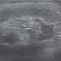
Acute pyelonephritis


Fig. 3.13


US: May be normal. Renal swelling, loss of corticomedullary differentiation, focal areas of abnormal echogenicity. Thickening of walls of pelvis/ureter and renal sinus hypoechogenicity.


Doppler: wedge-shaped areas of hypoperfusion.


DMSA: focal/diffuse decrease in tracer uptake.


Most commonly upper pole.


Imaging has limited role but may show structural abnormality that predisposes to infection or complications such as abscess.


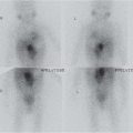
Acute obstructive nephropathy


US: variable dilatation of collecting system.


CT: may show calculus.


CECT/EU: persistent nephrogram and swollen parenchyma.


MRI: persistent nephrogram and delayed renal transit. MAG3: delayed renal transit time.


Causes: calculus, acute obstruction of partially obstructed system.


Nephroblastomatosis


Fig. 3.14


US: enlarged kidney with hypoechoic nodules and loss of corticomedullary differentiation.


CECT/MRI: Low attenuation nodules with poor enhancement. Predominantly subcapsular. Can be diffuse.


Usually bilateral.


Neonate to 2 y. Increased risk of Wilms tumor, especially with Beckwith-Wiedemann syndrome. Requires follow-up but can spontaneously regress.


Mesoblastic nephroma


Fig. 3.15a, b


US: predominantly solid hypoechoic/mixed echogenicity lesion.


Doppler: may show anechoic vascular ring around tumor.


CT: enhances postcontrast but less than normal renal parenchyma.


Cysts, hemorrhage, and necrosis rarely found.


Most common neonatal solid tumor. Ninety percent of patients are aged < 1 y.


Although benign, can rarely metastasize to lungs, brain, and bone.


Wilms tumor


US/CT/MRI: heterogeneous mass. Inferior vena cava and renal vein may show thrombus. Does not encase vessels. Nodal metastases. Low signal on T1-weighted and high on T2-weighted MRI. MRI most sensitive for caval patency.


Eighty percent occur before 5 y of age.


Four to thirteen percent are bilateral; 1% are familial. Five to ten percent have calcifications.


Associated with cryptorchidism, hemihypertrophy, hypospadias, sporadic aniridia.


Multilocular cystic renal tumor


US/CT/MRI: Well-demarcated cystic intrarenal mass. Septae enhanced. Cysts may show evidence of hemorrhage or protein on MRI.


Two age peaks: 3 mo to 4 y (boys > girls). Adults (mainly women).


Benign.


Autosomal dominant polycystic kidney disease (ADPKD)


Fig. 3.16a, b


See Table 3.6


May be unilateral initially in young patients.


Renal transplant rejection


US: Increased echogenicity. Loss of corticomedullary differentiation.


Doppler: decreased blood flow and increased resistive indices.


Acute tubular necrosis has a similar appearance.


Medullary sponge kidney


Fig. 3.17


(see bilateral medullary sponge kidneys, Table 3.6 )


Xanthogranulomatous pyelonephritis


Fig. 3.18a, b


US/CT: Calcified nodule in otherwise normal kidney seen in localized form. Diffuse form shows large lesion replacing entire kidney with loss of corticomedullary differentiation, cystic/necrotic areas, calcification/stones around renal pelvis.


Severe atypical, chronic renal parenchymal infection. Multiple causes: 70% stones. Diff cult to differentiate from tumor.


Acute renal vein thrombosis


Fig. 3.19


US: Enlarged, hypoechoic kidney with hyperechoic streaks. Loss of corticomedullary differentiation. Thrombus not always visible.


Doppler: Lack of flow in renal vein. Absent/reversed end-diastolic flow in intrarenal arteries.


MRI: most sensitive if US equivocal.


May be bilateral. On the left, may be associated with adrenal hemorrhage.


Causes: asphyxia, shock, dehydration, infant of diabetic mother.

Fig. 3.9 Duplex collecting system with dilatation of the upper moiety.
Fig. 3.10 Ureterocele. US image of the urinary bladder shows the presence of an ureterocele in the same patient as in Fig. 3.9 .
Fig. 3.11a, b Enlarged right kidney. (a) US image showing enlarged right kidney with altered axis; the left renal bed was empty. (b) MRI coronal postgadolinium image shows crossed fused renal ectopia of the left kidney.
Fig. 3.12a, b Focal contour defect. (a) US image of a focal contour defect in the right kidney from laceration caused by blunt abdominal trauma. (b) CECT image in the same patient defines the extent of laceration along with perinephric hematoma well.
Fig. 3.13 Enlarged kidney. US image of the right kidney in a child presenting with fever and urinary sepsis shows enlarged kidney with heterogeneously altered echotexture of the kidney. The normal corticomedullary distinction is somewhat lost.
Fig. 3.14 Nephroblastomatosis. CECT axial image in a child with nephroblastomatosis. Multiple hypoechoic nodules in both kidneys. The right kidney shows transformation of one of the rests into Wilms tumor.
Fig. 3.15a, b Mesoblastic nephroma. (a) Pre- and (b) postcontrast axial CT images show a mesoblastic nephroma in a neonate. The mass is hypodense and enhances heterogeneously with contrast.
Fig. 3.16a, b ADPKD. US images of bilaterally enlarged kidneys with multiple cysts in a child with ADPKD.
Fig. 3.17 Bilateral renal calcifications with dilated tubules on the EU.
Fig. 3.18a, b Xanthogranulomatous pyelonephritis. (a) US: markedly enlarged right kidney, focal alterations in echogenicity, calculi (arrow). (b) Precontrast CT: marked hydronephrosis on the left and intrarenal calcification.
Fig. 3.19 Acute renal vein thrombosis. Markedly enlarged kidney (arrows) with areas of echogenic hemorrhage.


Unilateral Renal Enlargement with Pelvicaliceal Dilatation


Hydronephrosis can be obstructive, nonobstructive, or secondary to reflux. The degree of dilatation depends predominantly on duration of reflux or obstruction and less on severity and can be assessed well on US. The presence of a dilated ureter helps determine cause. However, US is unable to assess severity of obstruction and function. Technetium-99m (99m Tc) mercaptoacetyltriglycine scintigraphy with Lasix washout is able to assess the degree of obstruction and remaining renal function, but if function is reduced in both kidneys, then erroneously “normal” results can be obtained. MRI is increasingly being used and is able to assess level and cause of hydronephrosis as well as function of kidneys, including GFR.


Table 3.5 Unilateral renal enlargement with PC dilatation

Diagnosis


Findings


Comments


Ureteropelvic junction (UPJ) obstruction


Fig. 3.20a–c


US: Dilatation of renal pelvis and calyces with varying parenchymal loss. Anteroposterior renal pelvis diameter > 10 m.


MAG3: Test of choice. Delayed excretion and retention of counts in pelvis. Activity curves allow quantitation of obstruction. Also allows indirect VCUG for reflux.


MRI: With gadolinium-diethylene triamine pentaacetic acid. Allows activity curves similar to NM as well as split renal function, GFR, and anatomic detail.


Associated with VUR (14%, which can be shown by VCUG), horseshoe kidney, lithiasis.


Ten to thirty percent are bilateral. Usually obstruction is partial.


Often diagnosed on prenatal US.


Hydronephrosis due to VUR


Dilated collecting system (see bilateral renal enlargement with PC dilatation, Table 3.7 )


Nonobstructive hydronephrosis or status postsurgically repaired urethral obstruction


US: hydronephrosis.


MAG3: normal washout of tracer with diuretic.


Megacalycosis/polycalycosis


EU: Polygonal multifaceted appearance of 15+ calyces.


Cortical thinning.


MAG3: no evidence of obstruction.


May be associated with primary megaureter.


At risk for lithiasis, infection. Thought to be secondary to underdevelopment of renal pyramids.


Multicystic dysplastic kidneys (MCDK)


Fig. 3.1, p. 240


Fig. 3.2, p. 240


US: Multiple large and some small noncommunicating cysts. Echogenic dysplastic tissue between cysts. No normal renal parenchyma.


NM: no renal excretion of isotope.


Associated with abnormal or atretic ipsilateral ureter. Ipsilateral genital abnormalities in 50% (e.g., cystic dysplasia of the rete testis).


Thirty percent of contralateral kidneys/ureter abnormal.


Distal ureteral stenosis


Variable dilatation of ureters and collecting system.


Often with minimal renal enlargement.


Associated with renal dysplasia.

Fig. 3.20a–c UPJ obstruction. (a) US image of dilated renal pelvis and hydronephrosis. (b) IVU shows persistent hydronephrosis after intravenous Lasix. (c) MAG3 scan postintravenous Lasix shows tracer retention in the renal PC system with delayed clearance in keeping with UPJ obstruction.

Only gold members can continue reading. Log In or Register to continue

Stay updated, free articles. Join our Telegram channel

Jul 12, 2020 | Posted by in PEDIATRIC IMAGING | Comments Off on Anomalous Renal Size

Full access? Get Clinical Tree

Get Clinical Tree app for offline access