Masses can be cystic (Table 3.25) or solid (Table 3.26). An adrenal mass in a fetus is usually a neuroblastoma, but after birth adrenal hemorrhage becomes more common (Figs. 3.68and 3.69). Initially, the postnatal appearance can be similar, but adrenal hemorrhage changes faster than a neuroblastoma; as most neonatal neuroblastomas involute spontaneously, follow-up US will help distinguish. In long-standing cases, the adrenal hemorrhage becomes calcified, and occult adrenal hemorrhage is probably the major cause of so-called idiopathic adrenal calcification (Fig. 3.70). In cases where there is still concern of neuroblastoma, especially in older children, US, CT, MRI, or NM may be very useful (Figs. 3.72, 3.73, 3.74, and 3.75). A rare differential diagnosis is an intra-abdominal sequestration.
Other tumors can occur in older children, the most common being pheochromocytoma, which is often predominately cystic and benign (Fig. 3.71a–c). CT and MRI are the most common modalities used to investigate these, although NM and positron emission tomography can be useful.
Fig. 3.67a, b Normal adrenal gland in neonates. Excellent differentiation is seen between the cortex and medulla in an adrenal gland with a Z configuration (a) and in an adrenal gland with an elongated configuration in a neonate with an absent kidney (b).
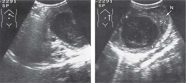
On US, echogenic, often hypoechoic, mass, with cystic change as resorption occurs over many weeks. Calcification frequently occurs later.
CT may demonstrate increased attenuation but this does aid differentiation from neuroblastoma.
Nearly all related to perinatal asphyxia. The hemorrhage can affect all or part of the gland and is bilateral in 10%. Rare in older children and is associated with severe blunt injury.
Adrenal cysts
These rare cysts can be confused with the upper pole of the kidney and should not be confused with an upper pole renal cyst, hydrocalyx, or hydronephrotic upper pole of a duplex kidney.
Caused most commonly by prenatal or postnatal hemorrhage. Microcysts are associated with Beckwith-Wiede-mann syndrome.
Hypoechoic, partly solid, mass with small cysts on US. CT and MRI show cystic and solid mass, with significant contrast enhancement. Iodine-123-metaiodobenzylguani-dine (123I-MIBG) scintigram: positive in 80% of cases.
Tumors originate from the medulla (70%) or sympathetic ganglia. Catecholamines are elevated and there may be associated endocrinopathies. In children, usually are benign. Bilateral in 24%.
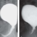
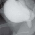
Fig. 3.68 Adrenal hemorrhage. The adrenal hemorrhage is cystic on US.Fig. 3.69 Adrenal hemorrhage. Postmortem CT of 30-week fetus following fetal death in utero: incidental right adrenal hemorrhage. (Image kindly provided by Dr. Michelle Fink, Director of Medical Imaging, Royal Melbourne Children’s Hospital, Australia.)Fig. 3.70 Idiopathic adrenal calcification. Unilateral right adrenal calcification is clearly seen in this plain film.Fig. 3.71a–c Right pheochromocytoma. There is a cystic lesion replacing most of the right adrenal gland shown on US (a), CT (b), and MRI (c).
Usually hyperechoic on US with calcification common. Lymph node metastases are common. Radiography, CT, and MRI show enhancing mass ± calcification. The primary tumor and metastases show uptake of 123I-MIBG scintigraphy
Urinary catecholamine levels are elevated. Seventy-five percent are intra-abdominal with majority in the adrenal. Usually present < 5 y old with peak in third year and up to 60% present with metastases.
Paraneoplastic syndromes, such as opsoclonus-myoclonus encephalopathy and intractable diarrhea, occur secondary to hormone release.
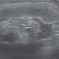
Intra-abdominal sequestration
US shows an echogenic mass adjacent to the adrenal gland.
Rare intra-abdominal mass may be mistaken for a neuroblastoma or adrenal hemorrhage but does not show any change over time unlike neuroblastoma and adrenal hemorrhage.
Ganglioneuroma
On cross-sectional imaging, these large solid tumors are usually homogeneous (except for calcification) and may show pressure on adjacent structures such as the ribs.
These slow growing benign tumors are more common and present later than neuroblastoma.
Rare tumors appear as solid masses on cross-sectional imaging.
Adenoma, adenocarcinoma that presents with Cushing syndrome (rare < 7 y), or endocrine malfunction, virilization.
Fig. 3.72 Neuroblastoma. US in a 1-year-old girl with right-sided abdominal mass shows a solid mass in the right side of the abdomen with fine speckled hyperechoic calcifications, which were confirmed to be a neuroblastoma.Fig. 3.73a, b Neuroblastoma. There is a large mass anterior to and infiltrating the hilum of the kidney on CECT. Note the characteristic surrounding of the aorta by the mass. The right (a) and left (b) renal arteries are also encased by tumor.Fig. 3.74 Neuroblastoma. There is a large mass of moderately high signal intensity arising from the left but crossing the midline and surrounding the aorta on T2-weighted MRI in a different patient from Fig. 3.73.Fig. 3.75 Neuroblastoma.123I-MIBG scintigram: an 8-year-old boy with an upper abdominal mass on CT scan, and avid tumor uptake on 123I-MIBG, that was proven to be adrenal at biopsy. (Image kindly provided by Dr. Arvind Kumar Sinha, Consultant Radiologist, Department of Diagnostic Imaging, National University Hospital, Singapore.)
Only gold members can continue reading. Log In or Register to continue