10.1055/b-0034-87882
The Adrenal Glands
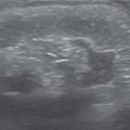
The adrenal glands are Z-shaped ( Fig. 3.67a ) unless there is absence or ectopia of the adjacent kidney, in which case they are discoid ( Fig. 3.67b ). They are large in the fetus and immediately after birth (see Fig. 3.67a, b ). On sonography, they are divided into two distinct hyperechoic and relatively hypoechoic layers: the cortex, and medulla (see Fig. 3.67a, b ). Physiologic involution of the adrenal glands begins after the first few weeks of life, ending by the fourth month. In this time, sonography is the modality of choice and can differentiate mass lesions. After this period, CT and MRI are more useful with MRI preferred, as it has no ionizing radiation.
Related posts:


Stay updated, free articles. Join our Telegram channel

Full access? Get Clinical Tree


