Chapter 92
Antrochoanal Polyp
Epidemiology
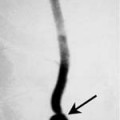
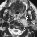
Antrochoanal polyp (ACP) is a polyp that originates from the maxillary antrum and extends into the nasal fossa usually through the secondary ostium of the maxillary sinus. ACP comprises about 5% of all polyps and most often presents in adolescents and young adults. These lesions are typically unilateral, but bilateral lesions have been reported in up to 30 to 40% of cases. The pressure of ACP are associated with allergies, systemic disorders which include multiple nasal polyposis, and cystic fibrosis.
Clinical Features
The most commonly presenting symptoms are nasal congestion and difficulty breathing. Patients may also present with sinus infection and drainage.
Pathology
A polyp is due to infiltration and expansion of the lamina propria of the schneiderian mucosa of the lining of the sinus or nasal cavity. Histologically, polyps are characterized by edema, proliferation of the connective tissue fibroblasts, and an inflammatory cellular infiltrate. An antrochoanal polyp is histologically indistinguishable from other forms of sinus or nasal polyps. In general, ACP tends to contain fewer eosinophils and mucous glands than nasal polyps.
Treatment
The treatment of choice is complete resection using a Caldwell-Luc or endoscopic intraantral approach. It is important that the stalk be resected. Incomplete removal of the stalk results in recurrent disease in 20 to 30% of cases.
Imaging Findings
Plain Films
There is typically unilateral opacification of the involved sinus without evidence of bone erosion.
CT