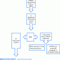
Location of arteriorgram
Catheter position
Contrast volume (ml)
Contrast time (s)
Image acquisitiona (frames/s)
Arch aortogram
Ascending aorta
30 ml
2
4
Innominate arteriogram
Innominate artery
18
3
3
Subclavian angiogram
Subclavian artery
12
3
3
Carotid angiogram
Common carotid artery
10
2
3
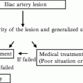
6 Management
Indications for treatment:
1.
Symptomatic disease
(a)
Cerebrovascular—ocular, hemispheric, or vertebrobasilar symptoms
(b)
Upper extremity ischemia
2.
Asymptomatic disease— >75 % stenosis
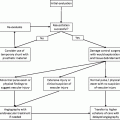
7 List of the Open Operative Choices
Transthoracic reconstruction
1.
Innominate artery disease
2.
Multivessel disease
Remote cervical reconstruction
1.
With single vessel occlusive disease
2.
Patients with prohibitive operative risk factors
8 Intervention: Techniques and Pitfalls
Prior to scheduling these patients, it is worthwhile to start them on ASA and Plavix (75 mg once a day) for 5 days. During the procedure they should receive intravenous heparin so as to maintain an activated clotting time between 250 and 300. The size of the stent is determined by the size of the blood vessel (Table 14.2).
Table 14.2
Vessel Size
Vessel | Size (mm) |
|---|---|
Innominate artery | 8–11 mm |
Subclavian artery | 6–8 mm |
Axillary artery | 5–7 mm |
Brachial artery | 5–7 mm |
8.1 Orificial Lesions
A.Subclavian Artery Lesions
1.
Occlusion: These are typically lesions which are difficult to recanalize. They usually extend from the origin of the subclavian artery to just proximal to the origin of the vertebral and the internal mammary arteries. Retrograde recanalization allows for more pushability to recanalize the occlusion
(a)
Access the brachial artery, preferably using a 4-Fr. micropuncture system under ultrasound visualization.
(b)
Femoral artery access is often useful using a 5-Fr. sheath and a pigtail catheter. Perform a flush aortogram to determine the location of the ostium of the artery as a target for the recanalization.
(c)
Exchange the brachial artery sheath to a to a 6-Fr. shuttle sheath (35–55 mm) over a 0.035″ non-hydrophilic wire.
(d)
Using a Kumpe catheter and a hydrophilic glide wire, attempt to recanalize the occluded lesion.
(e)
Change the wire to a stiff 0.014″ wire if the glide wire is unable to recanalize the lesion. This often requires the placement of a 0.014″ microcatheter (Quick Cross catheter, Spectranetics) for support.
(f)
Once the lesion has been recanalized, exchange the catheter for the dilator of the sheath and push the sheath forward across the lesion.
(g)
Introduce a balloon expandable stent (6–8 mm) once the sheath has traversed the lesion and the dilator removed. A balloon expandable stent is more appropriate for ostial lesions as they are more precise and also because they provide more radial force against these tough calcified lesions.
(h)
Position the stent at its appropriate location and pull back the sheath.
(i)
Deploy the stent and confirm position and patency
2.
Stenosis: These can typically be treated through a common femoral approach, but difficult arches (Type III) are sometimes easier to treat through the brachial approach.


(a)
Common femoral artery access is obtained.
(b)
A starter wire is placed over which the access needle is exchanged to a 6-Fr. sheath (35 cm).
(c)
Over the wire a pigtail catheter is placed and a flush aortogram is done.
(d)
Once the anatomy has been delineated, the appropriately shaped selective catheter (Kumpe/Simmons 1/Vitek) is preloaded through a guiding catheter (6 Fr.). These are loaded on the wire in exchange for the flush catheter, making sure that the wire has been exchanged to a stiffer variety for support.
(e)
The lesion is accessed.
Related posts:

Stay updated, free articles. Join our Telegram channel
Full access? Get Clinical Tree
