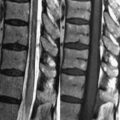
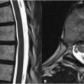
24 Arachnoid Cysts Arachnoid cysts are the most common congenital cystic brain entity, resulting from the failure to completely form an arachnoid villus or as sequelae of head trauma, leptomeningitis, or SAH. They form between the dura and arachnoid layers of the meninges or between the layers of the arachnoid. Along with berry aneurysms, they represent a frequent intracranial finding in polycystic kidney disease. Arachnoid cysts most commonly originate in the middle cranial fossa (Figs. 24.1A,B). Although typically much smaller than the lesion in Figs. 24.1A,B, arachnoid cysts are CSF-filled and thus characterized by isointensity to CSF on all MRI pulse sequences. This appearance includes high SI on T2WI (Fig. 24.1A), low SI on T1WI (Fig. 24.1B), and importantly low SI on FLAIR scans and DWI. Other characteristic locations include along the brain convexities and within the perimesencephalic cistern. Although typically asymptomatic, arachnoid cysts may become large enough to cause substantial mass effect on adjacent structures. Figure 24.1C demonstrates a large arachnoid cyst displacing the adjacent frontal lobe and also remodeling the inner table of the left frontal bone. Such erosions of the calvaria tend to be smooth. An expansile arachnoid cyst may grow large enough to obstruct the flow of CSF, resulting in ventricular dilatation, although this is quite rare. Retrocerebellar arachnoid cysts (Figs. 24.1D,E
![]()
Stay updated, free articles. Join our Telegram channel


Full access? Get Clinical Tree


