Presentation and Presenting Images
( ▶ Fig. 73.1, ▶ Fig. 73.2, ▶ Fig. 73.3, ▶ Fig. 73.4)
A 50-year-old female presents for routine screening mammography.
73.2 Key Images
73.2.1 Breast Tissue Density
The breasts are heterogeneously dense, which may obscure small masses.
73.2.2 Imaging Findings
The imaging of the right breast is normal (not shown). The left breast demonstrates possible architectural distortion (circle in ▶ Fig. 73.5 and ▶ Fig. 73.6), which may have been present on earlier based on comparison studies ( ▶ Fig. 73.3 and ▶ Fig. 73.4). and may not have appreciably changed. The possible 1.5-cm architectural distortion is in the upper inner quadrant at the 10 o’clock location, 8 cm from the nipple. The asterisks on the prior study ( ▶ Fig. 73.4) are computer-aided detection (CAD) marks unrelated to the possible architectural distortion. The circle marker (arrow) on the current study marks a skin mole.
73.3 BI-RADS Classification and Action
Category 0: Mammography: Incomplete. Need additional imaging evaluation and/or prior mammograms for comparison.
73.4 Diagnostic Images
( ▶ Fig. 73.7, ▶ Fig. 73.8, ▶ Fig. 73.9, ▶ Fig. 73.10, ▶ Fig. 73.11, ▶ Fig. 73.12, ▶ Fig. 73.13)
73.4.1 Imaging Findings
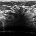
Digital breast tomosynthesis confirms the architectural distortion on slice 52 of 104 ( ▶ Fig. 73.7) on the left craniocaudal (CC) DBT movie and slice 26 of 98 ( ▶ Fig. 73.8) on the left lateromedial (LM) DBT movie. Retrospectively, after DBT, the finding can be seen on the left lateromedial mammogram ( ▶ Fig. 73.9). Ultrasound was performed and showed a corresponding area of architectural distortion (circle in ▶ Fig. 73.10 and ▶ Fig. 73.11). The lesion on ultrasound is hypoechoic with indistinct margins. A core needle biopsy was performed and postbiopsy mammograms showed adequate placement of the postbiopsy clip (box in ▶ Fig. 73.12 and ▶ Fig. 73.13).
73.5 BI-RADS Classification and Action
Category 4B: Moderate suspicion for malignancy
73.6 Differential Diagnosis
Ductal carcinoma in situ (DCIS) involving a radial scar: Architectural distortion is an uncommon presentation for DCIS. DCIS most commonly presents as microcalcifications. DCIS may be associated with radial scars, either involving the radial scar in this case or adjacent to a radial scar.
Radial scar: This finding was believed to have been present on the comparison mammogram and not appreciably changed, which makes a radial scar a reasonable possibility.
Summation artifact: The imaging finding persists on the DBT images and thus the imaging finding should not be dismissed.
73.7 Essential Facts
Mammography gives radiologists the opportunity to detect noninvasive breast cancer.
Ductal carcinoma in situ (DCIS) is noninvasive breast cancer: it is the proliferation of neoplastic cells of ducts of the breast without invasion of the parenchyma. The survival rate is almost 100% 10 years after diagnosis.
DCIS typically presents as calcifications. Sometimes mammography can “predict” the histology of DCIS. Linearly distributed calcifications are usually consistent with poorly differentiated DCIS; whereas calcifications with amorphous morphology are usually consistent with well-differentiated DCIS.
Radial scars have been shown to have a variable appearance on conventional mammography, presumably due to their planar configuration. The ability of DBT to improve lesion conspicuity reduces the effects of the planar configuration and allows detection of the radial scars.
Many radial scars will be visible on sonography and, when visible, may be indistinguishable from breast cancer. Radial scars are often more conspicuous on sonography than on mammography. When radial scars are visible on only one mammographic view and cannot be localized with certainty, sonography can be helpful to further evaluate these lesions. Now, that DBT can accurately localize one-view findings, it can better guide targeted ultrasound examinations.
The management and treatment of radial scars is controversial and varies from institution to institution.
73.8 Management and Digital Breast Tomosynthesis Principles
DBT acquires multiple images of the breast at multiple angles. The individual images are reconstructed into a series of thin slices typically 1 mm thick, which can be viewed in the same manner as computed tomography (CT) or magnetic resonance (MR) images, as single slices or as a dynamic cine on the workstation.
The ability to view images as a single slice or in dynamic mode eliminates the effects of overlapping breast tissue, which allows for better visualization and characterization of masses and architectural distortions.
The three-dimensional (3D) images of DBT eliminate the overlapping breast tissue, making lesions more conspicuous. In this case, the architectural distortion suspected on conventional mammographic imaging was confirmed on DBT.
Although it was not done in this case, DBT postbiopsy imaging could have been performed to further confirm that the architectural distortion was biopsied. Surgical excision was performed and no DCIS was identified in the surgical specimens.
73.9 Further Reading
[1] Cohen MA, Sferlazza SJ. Role of sonography in evaluation of radial scars of the breast. AJR Am J Roentgenol. 2000; 174(4): 1075‐1078 PubMed
[2] Ikeda DM, Andersson I. Ductal carcinoma in situ: atypical mammographic appearances. Radiology. 1989; 172(3): 661‐666 PubMed
[3] Leonard GD, Swain SM. Ductal carcinoma in situ, complexities and challenges. J Natl Cancer Inst. 2004; 96(12): 906‐920 PubMed

Fig. 73.1 Left craniocaudal (LCC) mammogram.
Stay updated, free articles. Join our Telegram channel

Full access? Get Clinical Tree


