Presentation and Presenting Images
A 71-year-old female presents for screening mammography.
55.2 Key Images
( ▶ Fig. 55.3, ▶ Fig. 55.4, ▶ Fig. 55.5, ▶ Fig. 55.6)
55.2.1 Breast Tissue Density
The breasts are heterogeneously dense, which may obscure small masses.
55.2.2 Imaging Findings
In the left breast at the 6 o’clock location in the anterior depth within the dense fibroglandular tissue, there is an area of architectural distortion. It is best seen on the tomosynthesis images ( ▶ Fig. 55.5 and ▶ Fig. 55.6), but can also be seen on the conventional digital mammograms ( ▶ Fig. 55.3 and ▶ Fig. 55.4).
55.3 BI-RADS Classification and Action
Category 0: Mammography: Incomplete. Need additional imaging evaluation and/or prior mammograms for comparison.
55.4 Diagnostic Images
( ▶ Fig. 55.7, ▶ Fig. 55.8, ▶ Fig. 55.9, ▶ Fig. 55.10, ▶ Fig. 55.11, ▶ Fig. 55.12, ▶ Fig. 55.13, ▶ Fig. 55.14, ▶ Fig. 55.15)
55.4.1 Imaging Findings
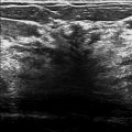
The diagnostic imaging nearly suggests that this area of architectural distortion is an artifact ( ▶ Fig. 55.7 and ▶ Fig. 55.8). However, on the CC spot-compression image ( ▶ Fig. 55.7 and ▶ Fig. 55.10), there appears to be a persistent finding. A targeted ultrasound was performed revealing a 7 × 7 × 10 mm irregular hypoechoic mass with indistinct margins and posterior acoustic shadowing at the 6 o’clock location, 1 cm from the nipple ( ▶ Fig. 55.11 and ▶ Fig. 55.12). This mass was biopsied and the ribbon biopsy clip is placed, as expected, in the location of the architectural distortion that was seen on the tomosynthesis images ( ▶ Fig. 55.13 and ▶ Fig. 55.14). This mass was eventually excised. The surgical specimen suggests a subtle mass with architectural distortion with the ribbon clip at the center ( ▶ Fig. 55.15).
55.5 BI-RADS Classification and Action
Category 4C: High suspicion for malignancy
55.6 Differential Diagnosis
Invasive lobular carcinoma (ILC): On image-guided biopsy and excision, this lesion was found to be a grade 1 ILC. This is a concordant diagnosis.
Invasive ductal carcinoma (IDC): There is significant overlap in the appearance of IDC and ILC on imaging. IDC would be a concordant diagnosis.
Radial scar: Radial scars are great mimickers of cancer. This would be concordant if the histopathologic size is similar to the imaging size. There is ongoing controversy about which radial scars need to be excised.
55.7 Essential Facts
Architectural distortion on mammography can be due to a malignant or a benign lesion.
Mammography alone often cannot distinguish between the etiology of architectural distortion without additional signs or information.
Malignancies that present as architectural distortion are typically IDC or ILC; however, ductal carcinoma in situ (DCIS) is also known to present as architectural distortion, but not as frequently.
Common benign causes of architectural distortion are radial scars, complex sclerosing lesions, and surgical scars (from excisions, lumpectomies, breast reductions, etc.).
Architectural distortion on screening mammography is less likely to represent malignancy compared to architectural distortion on diagnostic mammography.
Architectural distortion without a sonographic correlate is less likely to represent a malignancy.
55.8 Management and Digital Breast Tomosynthesis Principles
The addition of digital breast tomosynthesis (DBT) to full-field digital mammography (FFDM) improves the diagnostic performance of radiologists regardless of their level of experience.
The addition of DBT to FFDM increases the rate of cancer detection, reducing false-positives.
The unmasking effects of DBT reduces the underlying anatomical noise in the breast tissue. This is especially noted in dense breast tissue. In this case, the anatomical noise of the breast tissue easily obscures findings. The DBT images helped the reader to determine that the architectural distortion suggested on the craniocaudal (CC) FFDM was likely real and needed further evaluation with a diagnostic mammogram.
55.9 Further Reading
[1] Alakhras MM, Brennan PC, Rickard M, Bourne R, Mello-Thoms C. Effect of radiologists’ experience on breast cancer detection and localization using digital breast tomosynthesis. Eur Radiol. 2015; 25(2): 402‐409 PubMed
[2] Bahl M, Baker JA, Kinsey EN, Ghate SV. Architectural Distortion on Mammography: Correlation With Pathologic Outcomes and Predictors of Malignancy. AJR Am J Roentgenol. 2015; 205(6): 1339‐1345 PubMed

Fig. 55.1 Left craniocaudal (LCC) mammogram.
Stay updated, free articles. Join our Telegram channel

Full access? Get Clinical Tree


