Presentation and Presenting Images
A 74-year-old female presents for screening mammography.
77.2 Key Images
77.2.1 Breast Tissue Density
The breasts are almost entirely fatty.
77.2.2 Imaging Findings
The patient had conventional digital screening mammography. In the lower inner quadrant of the right breast at the 5 o’clock location, there is an area of architectural distortion (circle in ▶ Fig. 77.3 and ▶ Fig. 77.4). The mammogram of the left breast is normal (not shown).
77.3 BI-RADS Classification and Action
Category 0: Mammography: Incomplete. Need additional imaging evaluation and/or prior mammograms for comparison.
77.4 Diagnostic Images
( ▶ Fig. 77.5, ▶ Fig. 77.6, ▶ Fig. 77.7, ▶ Fig. 77.8, ▶ Fig. 77.9, ▶ Fig. 77.10, ▶ Fig. 77.11, ▶ Fig. 77.12, ▶ Fig. 77.13)
77.4.1 Imaging Findings
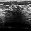
The diagnostic imaging ( ▶ Fig. 77.5, ▶ Fig. 77.6, ▶ Fig. 77.8, ▶ Fig. 77.9, and ▶ Fig. 77.11) demonstrates a persistent 1.5-cm area of architectural distortion located at the 5 o’clock location, which is also seen on the corresponding spot-compression digital breast tomosynthesis (DBT) images ( ▶ Fig. 77.7 and ▶ Fig. 77.10). An ultrasound was performed and no correlating abnormality was identified. The patient underwent stereotactic biopsy. The postbiopsy images demonstrate the biopsy clip at the location of the architectural distortion ( ▶ Fig. 77.12 and ▶ Fig. 77.13).
77.5 BI-RADS Classification and Action
Category 4B: Moderate suspicion for malignancy
77.6 Differential Diagnosis
Sclerosing adenosis: Sclerosing adenosis is the great mimicker of carcinoma. It can present as calcifications, a mass, and architectural distortion.
Carcinoma: Architectural distortion has a high positive predictive value (PPV) for carcinoma. This would be considered a concordant diagnosis for this lesion.
Radial scar: Radial scars typically present as an architectural distortion. This would be considered a concordant diagnosis for an image-guided biopsy provided that the histopathology expected for a radial scar is the dominant feature of the biopsy and not a small incidental finding.
77.7 Essential Facts
Sclerosing adenosis (SA) is a proliferation of the epithelium and myoepithelium that originates in the glandular lobules and is accompanied by desmoplasia.
SA can be associated with benign breast disorders, atypical lobular hyperplasia, and lobular carcinoma in situ.
SA can mimic carcinoma grossly and microscopically, and also on sonographic and mammographic imaging.
SA has variable appearances on imaging: calcifications, a mass, and architectural distortion. It is most commonly seen as calcifications. This case demonstrates a case of SA as architectural distortion.
77.8 Management and Digital Breast Tomosynthesis Principles
Architectural distortion seen on mammography has a high positive predictive value (PPV) for carcinoma (74.5%). Thus, it is important to sample the tissue. If the lesion is not seen on ultrasound (as in this case), then stereotactic biopsy or needle localization and excision should be performed to ensure that the lesion is not carcinoma.
DBT improves visualization of architectural distortion that is seen on conventional mammography, and in addition, identifies architectural distortion that is occult on conventional mammography.
The identification of occult architectural distortion by DBT accounts for the 12 to 45% of missed breast cancers.
DBT can improve the detection of architectural distortion; however, it cannot determine if the architectural distortion is of benign or malignant origin. A biopsy is required to make that determination.
77.9 Further Reading
[1] Bahl M, Baker JA, Kinsey EN, Ghate SV. Architectural Distortion on Mammography: Correlation With Pathologic Outcomes and Predictors of Malignancy. AJR Am J Roentgenol. 2015; 205(6): 1339‐1345 PubMed
[2] Günhan-Bilgen I, Memiş A, Ustün EE, Ozdemir N, Erhan Y. Sclerosing adenosis: mammographic and ultrasonographic findings with clinical and histopathological correlation. Eur J Radiol. 2002; 44(3): 232‐238 PubMed
[3] Partyka L, Lourenco AP, Mainiero MB. Detection of mammographically occult architectural distortion on digital breast tomosynthesis screening: initial clinical experience. AJR Am J Roentgenol. 2014; 203(1): 216‐222 PubMed

Fig. 77.1 Right craniocaudal (RCC) mammogram.
Stay updated, free articles. Join our Telegram channel

Full access? Get Clinical Tree


