Presentation and Presenting Images
( ▶ Fig. 53.1, ▶ Fig. 53.2, ▶ Fig. 53.3, ▶ Fig. 53.4)
A 56-year-old female presents for screening mammography.
53.2 Key Images
53.2.1 Breast Tissue Density
The breasts are heterogeneously dense, which may obscure small masses.
53.2.2 Imaging Findings
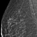
In the upper outer quadrant of the right breast at the 10 o’clock location in the posterior depth, there is a subtle architectural distortion. It is best seen on the tomosynthesis views ( ▶ Fig. 53.5 and ▶ Fig. 53.6). Once noted on the tomosynthesis views, the area can be faintly perceived on the conventional digital mammogram.
53.3 BI-RADS Classification and Action
Category 0: Mammography: Incomplete. Need additional imaging evaluation and/or prior mammograms for comparison.
53.4 Diagnostic Images
( ▶ Fig. 53.7, ▶ Fig. 53.8, ▶ Fig. 53.9, ▶ Fig. 53.10, ▶ Fig. 53.11, ▶ Fig. 53.12, ▶ Fig. 53.13, ▶ Fig. 53.14, ▶ Fig. 53.15, ▶ Fig. 53.16)
53.4.1 Imaging Findings
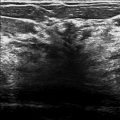
The diagnostic imaging does not offer much additional information about this area of architectural distortion ( ▶ Fig. 53.7, ▶ Fig. 53.8, and ▶ Fig. 53.9). The lateral view best identifies this area and suggests that it is about 1 cm in size ( ▶ Fig. 53.10). The anterior margin of the lesion creates an unnatural angle which makes it perceptible. A targeted ultrasound was performed because the findings seen on tomosynthesis suggested a mass associated with the architectural distortion and the overlying dense breast tissue obscured this finding on the conventional imaging. The ultrasound ( ▶ Fig. 53.11 and ▶ Fig. 53.12) reveals an irregular 1.5-cm mass with indistinct and angular margins with posterior acoustic shadowing at the 10 o’clock location, 5 cm from the nipple. Normal lymph nodes were seen in the ipsilateral axilla (not shown). This breast lesion was biopsied with ultrasound guidance and the postbiopsy mammogram demonstrates the ribbon clip ( ▶ Fig. 53.13 and ▶ Fig. 53.14) at the site of the initially questioned architectural distortion on the screening examination. A follow-up magnetic resonance (MR) image demonstrates that this is a singular lesion located in the breast with minimal background enhancement ( ▶ Fig. 53.15). The signal void from the biopsy clip can be seen in the mass on the sagittal MR image ( ▶ Fig. 53.16).
53.5 BI-RADS Classification and Action
Category 4C: High suspicion for malignancy
53.6 Differential Diagnosis
Invasive lobular carcinoma [ILC]: This very subtle lesion was difficult to detect even on DBT. The biopsy revealed a grade 2 ILC,. The MR image provides reassurance that there is only one lesion.
Invasive ductal carcinoma [IDC]: The imaging characteristics are not specific for ILC or IDC. They both would represent concordant results.
Fibroadenoma: Although this lesion is solid. The other imaging criteria including shape and margin characteristics on imaging should suggest a high suspicion of malignancy. A biopsy result of fibroadenoma would be discordant.
53.7 Essential Facts
Architectural distortion has a positive predictive value (PPV) of 74.5% on mammography. This study by Bahl and colleagues (2015) did not include digital breast tomosynthesis (DBT). They recognize that DBT may further improve the detection of architectural distortion.
Early studies have suggested that a diagnostic mammogram could be omitted for DBT findings other than those with calcifications. Brandt and colleagues (2013) found that in 93 to 99% of these cases the two-view screening DBT was adequate for mammographic evaluation.
Two additional similar studies by Poplack and Hakim found that DBT was equivalent or better to a conventional diagnostic mammogram in 89% and 81% of these cases, respectively.
In cases with calcifications, DBT has inferior image quality compared to a conventional diagnostic mammogram.
53.8 Management and Digital Breast Tomosynthesis Principles
Architectural distortion seen on a diagnostic mammogram was more likely to represent a malignancy than that detected on a screening mammogram. It is recognized that some screening findings will eventually be assessed to be benign on diagnostic work-up.
The implementation of DBT is evolving. Practices may differ from suggested guidelines. More research and experience is needed to determine the optimal imaging protocol.
53.9 Further Reading
[1] Bahl M, Baker JA, Kinsey EN, Ghate SV. Architectural Distortion on Mammography: Correlation With Pathologic Outcomes and Predictors of Malignancy. AJR Am J Roentgenol. 2015; 205(6): 1339‐1345 PubMed
[2] Brandt KR, Craig DA, Hoskins TL, et al. Can digital breast tomosynthesis replace conventional diagnostic mammography views for screening recalls without calcifications? A comparison study in a simulated clinical setting. AJR Am J Roentgenol. 2013; 200(2): 291‐298 PubMed

Fig. 53.1 Right craniocaudal (RCC) mammogram.
Stay updated, free articles. Join our Telegram channel

Full access? Get Clinical Tree


