Presentation and Presenting Images
A 75-year-old female presents for asymptomatic screening mammography.
71.2 Key Image
( ▶ Fig. 71.3)
71.2.1 Breast Tissue Density
There are scattered areas of fibroglandular density.
71.2.2 Imaging Findings
The patient had a conventional digital screening mammogram. There is architectural distortion possibly associated with calcifications in the lateral aspect in the middle depth on the craniocaudal (CC) view of the right breast ( ▶ Fig. 71.3). The mammogram of the left breast was normal (not shown).
71.3 BI-RADS Classification and Action
Category 0: Mammography: Incomplete. Need additional imaging evaluation and/or prior mammograms for comparison.
71.4 Diagnostic Images
( ▶ Fig. 71.4, ▶ Fig. 71.5, ▶ Fig. 71.6, ▶ Fig. 71.7, ▶ Fig. 71.8, ▶ Fig. 71.9, ▶ Fig. 71.10, ▶ Fig. 71.11, ▶ Fig. 71.12, ▶ Fig. 71.13, ▶ Fig. 71.14)
71.4.1 Imaging Findings
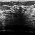
The architectural distortion could be reproduced on the CC spot-compression image ( ▶ Fig. 71.4 and ▶ Fig. 71.5). However, there was still confusion about the location in the lateral view. Thus, combination CC and mediolateral (ML) full-field digital mammogram (FFDM) with digital breast tomosynthesis (DBT) imaging was obtained ( ▶ Fig. 71.6, ▶ Fig. 71.7, ▶ Fig. 71.8, and ▶ Fig. 71.9). These images support an architectural distortion correlate in the lateral projection. There are scattered calcifications in the breast; however, there is a group of coarse heterogeneous calcifications that appear to follow the architectural distortion ( ▶ Fig. 71.10 and ▶ Fig. 71.11). This area was localized to the upper outer quadrant around the 10 o’clock location. An ultrasound was then performed.
The targeted ultrasound revealed a very subtle irregular hypoechoic mass with indistinct margins located at the 10 o’clock location, 9 cm from the nipple ( ▶ Fig. 71.12). This lesion was biopsied with ultrasound guidance. The clip on the postprocedure mammogram appears to be located at the sight of the architectural distortion ( ▶ Fig. 71.13 and ▶ Fig. 71.14). If the clip was not located at this site, it would be reasonable to biopsy this finding with stereotactic technique and use the calcifications as the target.
71.5 BI-RADS Classification and Action
Category 4B: Moderate suspicion for malignancy
71.6 Differential Diagnosis
Invasive ductal carcinoma (IDC): IDC can present as an architectural distortion with or without associated calcifications. Architectural distortion is not very common, but when present has a high predictive value for carcinoma. Biopsy of this lesion was grade 1 IDC with DCIS.
Ductal carcinoma in situ (DCIS) : The calcifications could be associated with DCIS. Invasive cancer is more likely to be associated with architectural distortion than DCIS. If DCIS is identified on image-guided biopsy, it is possible that an upgrade to invasive cancer could be found at surgical excision.
Sclerosing adenosis: Sclerosing adenosis is a great mimicker of carcinoma. If the lesion seen on imaging is comparable to the size of the lesion seen at pathology, it could be considered concordant.
71.7 Essential Facts
Mammographic features alone cannot be used to differentiate benign from malignant causes of architectural distortion.
The positive predictive value (PPV) of architectural distortion for cancer is 74.5%.
Architectural distortion with calcifications and without calcifications do not have significant differences in their rates of malignancy.
If the architectural distortion seen mammographically or on digital breast tomosynthesis (DBT) does not appear to have a sonographic correlate, the presence of calcifications can aid in targeting the biopsy with traditional stereotactic techniques.
Architectural distortion also can be biopsied with the new DBT-guided stereotactic biopsy.
Due to its high PPV for cancer, architectural distortion does not fit the criteria for observation.
71.8 Management and Digital Breast Tomosynthesis Principles
Architectural distortion and asymmetries seen on conventional mammography are often proven to be overlapping tissue on DBT.
Architectural distortion is less likely to be a malignancy if detected on screening mammography, and more likely if seen on diagnostic mammography.
Early studies suggest that architectural distortion seen on DBT without a sonographic correlate is more likely to be a radial scar than a malignancy. Further studies are needed to determine if architectural distortion seen only on DBT without an ultrasound correlate can be followed or requires biopsy.
The cancer detection rate for architectural distortion seen on DBT that is mammographically occult has been reported to be 21.1% and 47.2%.
71.9 Further Reading
[1] Bahl M, Baker JA, Kinsey EN, Ghate SV. Architectural Distortion on Mammography: Correlation With Pathologic Outcomes and Predictors of Malignancy. AJR Am J Roentgenol. 2015; 205(6): 1339‐1345 PubMed

Fig. 71.1 Right craniocaudal (RCC) mammogram.
Stay updated, free articles. Join our Telegram channel

Full access? Get Clinical Tree


