Presentation and Presenting Images
( ▶ Fig. 48.1, ▶ Fig. 48.2, ▶ Fig. 48.3, ▶ Fig. 48.4)
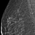
A 50-year-old female with a history of left breast cancer treated 8 years ago presents for screening mammography.
48.2 Key Images
48.2.1 Breast Tissue Density
The breasts are extremely dense, which lowers the sensitivity of mammography.
48.2.2 Imaging Findings
In the upper outer quadrant of the right breast, at the edge of the glandular tissue, there is a possible architectural distortion seen at the 10 o’clock position in the posterior depth. This is only appreciated on the digital breast tomosynthesis (DBT) images ( ▶ Fig. 48.5 and ▶ Fig. 48.6).
48.3 BI-RADS Classification and Action
Category 0: Mammography: Incomplete. Need additional imaging evaluation and/or prior mammograms for comparison.
48.4 Diagnostic Images
( ▶ Fig. 48.7, ▶ Fig. 48.8, ▶ Fig. 48.9, ▶ Fig. 48.10, ▶ Fig. 48.11, ▶ Fig. 48.12, ▶ Fig. 48.13, ▶ Fig. 48.14, ▶ Fig. 48.15, ▶ Fig. 48.16)
48.4.1 Imaging Findings
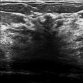
The diagnostic imaging identifies a 1.1-cm subtle focal asymmetry ( ▶ Fig. 48.7, ▶ Fig. 48.10 and ▶ Fig. 48.8, ▶ Fig. 48.11) at the edge of the glandular tissue at the 10 o’clock location with the help of the prior screening DBT ( ▶ Fig. 48.5 and ▶ Fig. 48.6). The targeted ultrasound reveals a 0.6 × 1.0 × 0.9 cm irregular hypoechoic mass with angular and indistinct margins located at 10 o’clock, 10 cm from the nipple ( ▶ Fig. 48.12 and ▶ Fig. 48.13). There were normal appearing right axillary nodes (not shown). The postbiopsy mammogram demonstrates the ribbon biopsy clip is the posterior upper outer quadrant in the expected location ( ▶ Fig. 48.14 and ▶ Fig. 48.15). Note that the patient does have a coarse calcification in the lateral aspect of the breast (not to be mistaken for the biopsy clip, which is smaller). Due to the patient’s prior history and underlying dense breast tissue, she underwent an MR examination. The MR image reveals this solitary enhancing right breast lesion in the upper outer quadrant at the periphery of the glandular tissue ( ▶ Fig. 48.16).
48.5 BI-RADS Classification and Action
Category 4C: High suspicion for malignancy
48.6 Differential Diagnosis
Carcinoma (invasive lobular carcinoma [ILC]): The initial difficulty with this case was detection. The DBT images revealed this lesion and the subsequent imaging, especially the ultrasound in the diagnostic evaluation, confirmed this lesion. This lesion is consistent with carcinoma.
Radial scar: Typically radial scars do not have a significant mass equivalent on ultrasound. If the diagnosis based on the biopsy of this lesion was a radial scar, to be concordant, the histopathologic size would have to match the imaging size. Otherwise, it would be discordant, and excision of the lesion would be recommended.
Fibroadenoma: Fibroadenomas have variable features. However, this lesion appears more suspicious, and, thus, a diagnosis of a fibroadenoma on image-guided biopsy would be considered discordant, and excision would be recommended.
48.7 Essential Facts
ILC accounts for 10 to 15% of invasive breast cancers.
ILC most frequently manifests mammographically as a mass (44–65% of cases) and secondly as an architectural distortion (10–34% of cases).
Ray and colleagues (2015) reported that DBT identified 7% of lesions recommended for biopsy that were occult on full-field digital mammography (FFDM). Over half of these mammographically occult lesions were malignant.
Architectural distortion appears to represent the most common finding seen on DBT that is occult on FFDM.
48.8 Management and Digital Breast Tomosynthesis Principles
There is a higher false negative rate for ILC (up to 19%) on mammography compared to other invasive breast cancers.
DBT improves the ability of detecting subtle distortion by reducing the effects of overlapping tissue.
DBT also allows the dismissal of pseudo–architectural distortion that is created by summation artifacts.
ILC is the most likely invasive breast cancer to be occult to imaging and physical examination. More studies are needed to see if DBT can have an impact on ILC detection.
Ray and colleagues’ (2015) limited retrospective study suggested that ILC had a higher frequency of detection with DBT, accounting for one-third of their identified cancers.
48.9 Further Reading
[1] Lopez JK, Bassett LW. Invasive lobular carcinoma of the breast: spectrum of mammographic, US, and MR imaging findings. Radiographics. 2009; 29(1): 165‐176 PubMed
[2] Ray KM, Turner E, Sickles EA, Joe BN. Suspicious Findings at Digital Breast Tomosynthesis Occult to Conventional Digital Mammography: Imaging Features and Pathology Findings. Breast J. 2015; 21(5): 538‐542 PubMed

Fig. 48.1 Right craniocaudal (RCC) mammogram.
Stay updated, free articles. Join our Telegram channel

Full access? Get Clinical Tree


