Presentation and Presenting Images
( ▶ Fig. 5.1, ▶ Fig. 5.2, ▶ Fig. 5.3, ▶ Fig. 5.4, ▶ Fig. 5.5, ▶ Fig. 5.6)
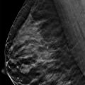
A 62-year-old female with a history of left mastectomy for invasive ductal carcinoma and right reduction mammoplasty presents for routine mammography.
5.2 Key Images
( ▶ Fig. 5.7, ▶ Fig. 5.8, ▶ Fig. 5.9, ▶ Fig. 5.10, ▶ Fig. 5.11)
5.2.1 Breast Tissue Density
The breasts are heterogeneously dense, which may obscure small masses.
5.2.2 Imaging Findings
There is no imaging of the left breast as the patient had a mastectomy. The right breast demonstrates architectural distortion centrally (circle in ▶ Fig. 5.7, ▶ Fig. 5.8, ▶ Fig. 5.10, and ▶ Fig. 5.11) and elevation of the nipple (arrow in ▶ Fig. 5.8 and ▶ Fig. 5.9).
5.3 BI-RADS Classification and Action
Category 2: Benign
5.4 Differential Diagnosis
Postsurgical changes: There is central architectural distortion and elevation of the nipple. These imaging findings correlate with the surgical changes of reduction mammoplasty.
Radial scar: Although a radial scar may present as architectural distortion, the clinical history is less appropriate for this diagnosis.
Breast cancer: Patients with a prior history of breast cancer are at increased risk for developing another cancer, but the findings in this case are most consistent with postsurgical changes.
5.5 Essential Facts
According to Danikas and colleagues (2001), the most common mammographic findings seen after reduction mammoplasty were parenchymal redistribution and elevation of the nipple. These authors felt that mammographic findings after reduction mammoplasty are predictable; therefore knowledge of these findings will prevent unnecessary biopsies and make the diagnosis of lesions unrelated to the procedure easier.
All patients over 35 years of age having reduction mammoplasty surgery should have a preoperative and a postoperative mammogram for future reference.
Postsurgical changes following reduction mammoplasty do not significantly hinder evaluation of screening mammograms and the detection of breast cancers.
5.6 Management and Digital Breast Tomosynthesis Principles
Obtaining the two-dimensional (2D) mammogram along with tomosynthesis allowed direct comparison between the 2D mammogram and tomosynthesis. Comparing the two studies in this example, the breast is smaller, the nipple position is higher, and there is central architectural distortion.
Tomosynthesis is helpful in identifying lesions unrelated to the reduction mammoplasty procedure and the postsurgical changes.
The process of reviewing the images acquired with tomosynthesis takes longer, but early studies suggest that the number of callbacks are reduced.
5.7 Further Reading
[1] Danikas D, Theodorou SJ, Kokkalis G, Vasiou K, Kyriakopoulou K. Mammographic findings following reduction mammoplasty. Aesthetic Plast Surg. 2001; 25(4): 283‐285 PubMed
[2] Muir TM, Tresham J, Fritschi L, Wylie E. Screening for breast cancer post reduction mammoplasty. Clin Radiol. 2010; 65(3): 198‐205 PubMed

Fig. 5.1 Right craniocaudal (RCC) mammogram.
Related posts:


Stay updated, free articles. Join our Telegram channel

Full access? Get Clinical Tree


