13 Arterial Anatomy of the Extremities
The evaluation of arterial disease of the extremities requires knowledge of vascular anatomy. This chapter provides this basic information for the upper and lower extremities. Normal anatomy, common variants, and major collateral routes1–6 are illustrated, primarily by representative arteriograms. The chapter is formatted as a series of captioned illustrations, with the bulk of the instructional material contained within the figure captions.
Upper Extremity
Normal Features
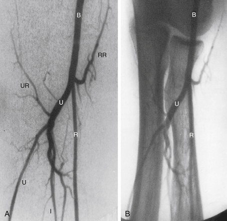
The normal arterial anatomy of the upper extremity is depicted graphically in Figure 13-1. Figures 13-2 to 13-5 are detailed arteriographic views of specific regions of the upper extremity arterial tree, beginning at the aorta and extending to the digits. These figures should be reviewed carefully, because their legends provide the instructional content.
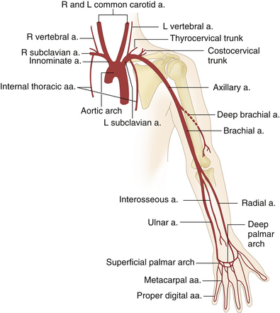
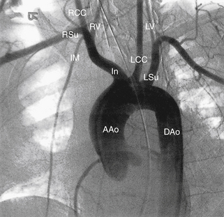
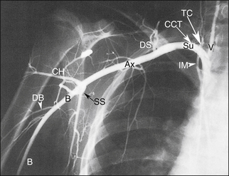
FIGURE 13-3 The subclavian artery (Su) becomes the axillary artery (Ax) at the lateral margin of the first rib. The axillary artery, in turn, becomes the brachial artery (B) after crossing the inferolateral margin of the teres major muscle.3 The thyrocervical (TC) and costocervical (CCT) trunks are noteworthy branches of the subclavian artery, because they may be mistaken for the vertebral artery (V) during duplex examination. The multiple branches that supply the scapular musculature serve as collaterals when the subclavian or innominate arteries are obstructed. CH, circumflex humeral artery; DB, deep brachial artery; DS, dorsal scapular artery; IM, internal mammary artery; SS, subscapular artery.
Anatomic Variants
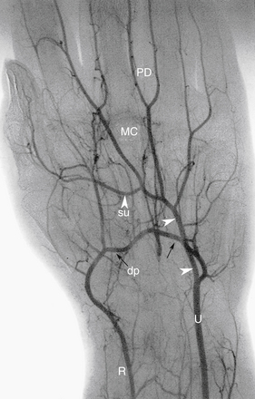
Many anatomic variants can occur in the arterial tree of the upper extremities. The more commonly encountered variants are presented in Table 13-1.1–3 Familiarity with these variants can prevent confusion and error during duplex examination. An example of an upper extremity anatomic variant is presented in Figure 13-6.
TABLE 13-1 Arterial Variants of the Upper Extremity
| Structure | Variant | Frequency of Occurrence in the Population (%) |
|---|---|---|
| Aortic arch and great vessels | Common origin of the right brachiocephalic and left common carotid arteries | 22 |
| Left vertebral artery origin directly from the aorta | 4-6 | |
| Common origin of both common carotid arteries | <1 | |
| Arm and forearm | Radial artery origin from the axillary artery | 1-3 |
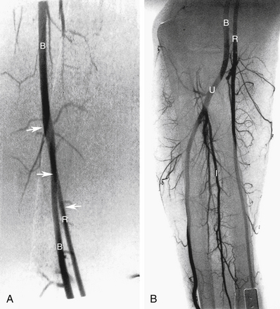
| Early division of the brachial artery: 1. High origin of the radial artery (Figure 13-6) 2. Accessory (duplicated) brachial artery | 19 | |
| Ulnar artery origin from the brachial or axillary artery | 2-3 | |
| Low origin (5-7 cm below elbow joint) of ulnar artery | <1 | |
| Persistent median artery | 2-4 |
Collateral Routes
Many of the tributaries seen in Figures 13-1 to 13-6 may serve as collaterals when the main arterial trunks of the upper extremity are blocked.
The following is a summary of the more common collateral routes.2
1. Obstruction of the proximal subclavian or brachiocephalic arteries
Related posts:
 Normal Cerebrovascular Anatomy and Collateral Pathways
 Controversies in Venous Ultrasound
 Nonimaging Physiologic Tests for Assessment of Lower Extremity Arterial Disease
 Duplex Ultrasound Evaluation of the Uterus and Ovaries
 Extremity Venous Anatomy and Technique for Ultrasound Examination
Stay updated, free articles. Join our Telegram channel

Full access? Get Clinical Tree


