, Marilyn J. Siegel2, Tomasz Miszalski-Jamka3, 4 and Robert Pelberg1
(1)
The Christ Hospital Heart and Vascular Center of Greater Cincinnati, The Lindner Center for Research and Education, Cincinnati, OH, USA
(2)
Mallinckrodt Institute of Radiology, Washington University School of Medicine, St. Louis, Missouri, USA
(3)
Department of Clinical Radiology and Imaging Diagnostics, 4th Military Hospital, Wrocław, Poland
(4)
Center for Diagnosis Prevention and Telemedicine, John Paul II Hospital, Kraków, Poland
Abstract
Arterial switch (Jatene) procedure, which was developed in the late 1970s, is currently the method of choice for treatment of transposition of the great arteries (TGA). It reestablishes the left ventricle as a systemic pump, avoiding systemic ventricular failure associated with the atrial switch operation. This procedure is indicated for patients with TGA with an intact ventricular septum or with a small ventricular septal defect (VSD). Patients with large nonrestrictive VSDs, left ventricular outflow obstruction, or increased pulmonary vascular resistance may not be candidates for the arterial switch procedure.
Arterial switch (Jatene) procedure, which was developed in the late 1970s, is currently the method of choice for treatment of transposition of the great arteries (TGA). It reestablishes the left ventricle as a systemic pump, avoiding systemic ventricular failure associated with the atrial switch operation. This procedure is indicated for patients with TGA with an intact ventricular septum or with a small ventricular septal defect (VSD). Patients with large nonrestrictive VSDs, left ventricular outflow obstruction, or increased pulmonary vascular resistance may not be candidates for the arterial switch procedure.
The Jatene procedure is typically performed in the first 2 weeks of life. Following aortic cross-clamping, the left and right coronary artery ostia and a cuff of the adjacent aortic wall attached as “buttons” are excised from the aorta, and the proximal sections of the coronary arteries are separated from the surface of the heart. This technique prevents distortion after anastomosis to the neoaorta. Usually, the coronary implantation sites are located at the left and right anterior positions at the base of the neoaorta. Next the aorta is transected above the coronary artery ostia and the pulmonary artery is transected above the pulmonic valve.
Subsequently, a Lecompte maneuver (added in the 1980s) is performed and involves positioning the distal pulmonary artery and its branches anterior to the aortic root. This maneuver maximizes the length of the aorta, thus further reducing the risk of coronary artery kinking and stenosis. The distal ascending aorta is now anastomosed to the “new” aortic root (neoaorta) and the pulmonary artery is anastomosed to the new pulmonic root (neo-pulmonary artery). The coronary arteries are then shifted posteriorly and reimplanted into the facing sinuses of the neoaorta just above its origin from the morphologic left ventricle (Figs. 24.1 and 24.2). Because of the potential complication of stenosis at the anastomotic site of the pulmonary artery to the morphologic right ventricle, reconstruction of the pulmonary artery is often undertaken utilizing a pericardial patch or cryopreserved pulmonary artery homograft patch. Any VSD is also repaired at the time of surgery.

Get Clinical Tree app for offline access
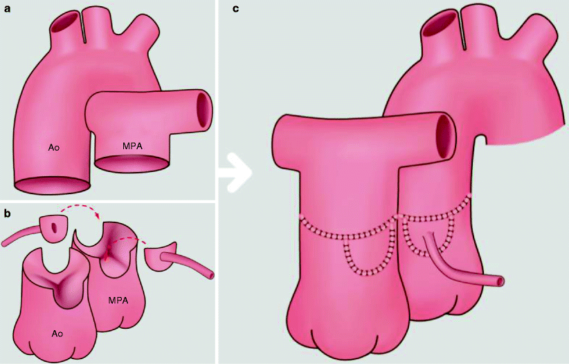
Fig. 24.1
Arterial switch operation. Reattachment of the great arteries to the contralateral ventricles with reimplantation of the coronary artery ostia into the neoaorta. Panel (a) shows the unrepaired transposition morphology prior to surgery. The aorta is anterior and to the right of the pulmonary artery. Panel (b) depicts the Jatene procedure where the coronary ostia and a “button” of surrounding aortic wall are excised from the aorta and the proximal sections of the coronary arteries are separated from the heart, which prevents distortion after anastomosis to the neoaorta. Panel (c) shows the aorta transplanted onto the pulmonary root. The great arteries are arranged using the Lecompte maneuver, with the pulmonary artery positioned anterior to the ascending aorta. Ao aorta, MPA main pulmonary artery
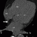
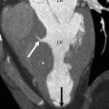
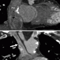
Fig. 24.2
Jatene arterial switch. A 22-year-old woman with transposition of the great arteries status post surgical repair and reimplantation of a single right coronary artery. Panel (a) axial and (b) sagittal images show the main pulmonary artery (MPA) anterior to the aorta (Ao). The right and left pulmonary arteries drape over the ascending aorta. There is no stenosis of the right or the left pulmonary artery. Note the parallel orientation of the aorta and pulmonary artery. Panel (c) is an axial view in a more caudal plane showing the pulmonic valve (P) anterior to the aortic valve (A). Panels (d) and (e) are reformatted scans showing a single, reimplanted coronary artery (arrows) originating above the sinotubular junction from the right coronary cusp of the aortic valve. The origin of the single coronary artery is immediately posterior to the main pulmonary artery (MPA) and anterior to the aorta (Ao


Related posts:


Stay updated, free articles. Join our Telegram channel
Full access? Get Clinical Tree
