35 Arteries of the Forearm
B. Meyer, L. Sonnow
Fig. 35.1 summarizes all cases in which the arteries of the forearm follow a “normal” course, leaving the origin of these arteries out of consideration. Fig. 35.2, Fig. 35.3, Fig. 35.4, and Fig. 35.5 demonstrate the embryological basis for the more important variations. The origin of an ulnar artery as a branch of the superficial brachial artery running parallel to a brachial artery (Section 34.3) has not been described. The superficial arteries of the forearm usually branch from a superficial brachial artery, which runs parallel to the brachial artery (Section 34.3). Sometimes the superficial arteries of the forearm are branches of the brachial artery.1–9
35.1 “Normal” Arterial Pattern of the Forearm (Radial, Ulnar, and Interosseous Artery) (84%)
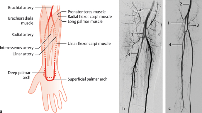
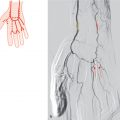
Fig. 35.1 Normal arterial pattern of the forearm. Schematic (a) and DSA images of the forearm from two different patients (b,c). 1 Radial artery; 2 brachial artery; 3 ulnar artery; 4 interosseous artery.

Fig. 35.2 All arteries of the forearm branch from the branchial artery (70%). See also Section 34.1. Schematic.
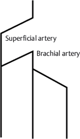
Fig. 35.3 All arteries of the forearm are branches of a superficial branchial artery (6%). Schematic.
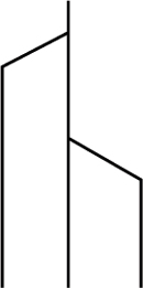
Fig. 35.4 The radial artery originates from the superficial branchial artery, and the ulnar and interosseous artery from the branchial artery (5%). Schematic.

Fig. 35.5 Same as Fig. 35.3, but with a large anastomosis between the brachial and radial arteries in the cubital region (3%). Schematic.
35.2 Superficial Arteries of the Forearm (Superficial Antebrachial Artery) (8%)
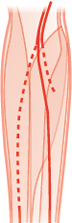
Fig. 35.6 Superficial ulnar artery replaces the ulnar artery (3%). Schematic.
Related posts:


Stay updated, free articles. Join our Telegram channel

Full access? Get Clinical Tree


