9 Inferior Phrenic Arteries
K.I. Ringe
These small arteries supply the diaphragm from the abdominal side. They anastomose with the small superior phrenic arteries, branches of the thoracic aorta, and the musculophrenic and the pericardiophrenic arteries, which are branches of the internal thoracic arteries. The inferior phrenic arteries stem from the mesonephric arteries and thus also supply the adrenal gland via the superior suprarenal artery. This embryologic development elucidates the rare origins of the inferior phrenic arteries from the renal, testicular, or ovarian arteries (Fig. 9.7, Fig. 9.9). Their origin from the celiac trunk (Fig. 9.8) has been found surprisingly often and can only be explained by a secondary transposition on account of the vessels’ different growth rates. The normal pattern described in textbooks accounts for only about a quarter of all cases.1–12
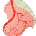
9.1 Both Inferior Phrenic Arteries Originate from a Common Trunk (33%)

Fig. 9.1 Origin from the aorta just above the celiac trunk (18%). Schematic (a) and CT images (b,c). MIP, axial (b) and sagittal (c) views. 1 Right phrenic artery; 2 left phrenic artery; 3 common phrenic branch; 4 celiac trunk; 5 superior mesenteric artery.

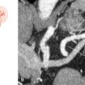
Fig. 9.2 Origin from the celiac trunk (14%). Schematic (a) and CT images (b,c). AvIP, coronal view (b); and MIP, sagittal view (c). A transjugular intrahepatic portosystemic shunt (TIPS) can be appreciated. 1 TIPS; 2 left phrenic artery; 3 superior mesenteric artery; 4 celiac trunk; 5 right phrenic artery; 6 common hepatic artery; 7 phrenic artery.

Fig. 9.3 Origin from the left gastric artery (1%). Schematic (a) and axial MIP CT at the level of the celiac trunk (b). 1 Celiac trunk; 2 left gastric artery; 3 splenic artery; 4 left phrenic artery; 5 right phrenic artery; 6 common hepatic artery.
9.2 The Inferior Phrenic Arteries Have Separate Origins (67%)

Fig. 9.4 Origin from the abdominal aorta (right side 29%; left side 24%). Schematic (a), axial MIP CT above the level of the celiac trunk (b), and coronal VR CT (c). In c an upper polar renal artery can be appreciated on the left (*). 1 Right phrenic artery; 2 left phrenic artery; 3 splenic artery; 4 celiac trunk; 5 superior mesenteric artery.
Related posts:


Stay updated, free articles. Join our Telegram channel

Full access? Get Clinical Tree


