13 Celiac Trunk
K.I. Ringe
Of the three main branches of the celiac trunk, the common hepatic artery has the highest frequency of anomalies.1 For details of anomalies of the hepatic, left gastric, and splenic artery, see Chapters 14, 16, and 17. When a complete trunk is described (Fig. 13.1), this does not mean that the common hepatic artery has all “normal” branches, but only that a major part of the common hepatic artery, for example, the gastroduodenal artery, arises from the celiac trunk. An accessory hepatic artery from the superior mesenteric or left gastric artery was not included in this chapter. As in Fig. 13.10 (hepatomesenteric trunk), only those cases have been included in which no major part of the common hepatic artery comes from the celiac trunk. A clear-cut differentiation between the types shown in Fig. 13.1 and Fig. 13.2 is not always possible. The most frequent accessory branch is the inferior phrenic artery (in ~50%; see Chapter 9). In this chapter, only variations of the typical branches of the celiac trunk are included.2–19
The origins from the aorta of the celiac trunk and the superior mesenteric artery are close to each other and they quite often form common trunks.18,20 For the sake of clarity, the origins are drawn further apart than is the case in situ.
13.1 Complete Celiac Trunk (Gastrohepatolienal Trunk) (84%)

Fig. 13.1 “Normal” type as seen in textbooks: the celiac trunk is formed by the left gastric common hepatic and splenic artery (tripus Halleri) (~25%). Schematic (a) and VR CT image (b) in a patient with portal hypertension and status post transjugular intrahepatic portosystemic shunt implantation. The CTA demonstrates a normal branching pattern of the celiac trunk. 1 Transjugular intrahepatic portosystemic shunt; 2 left gastric artery; 3 celiac trunk; 4 splenic artery; 5 common hepatic artery.

Fig. 13.2 The celiac trunk leaves the left gastric artery as a side branch and divides more distally into the common hepatic and the splenic artery (~49%). Schematic (a), coronal VR CT (b), and sagittal MIP CT (c). 1 Left gastric artery; 2 celiac trunk.

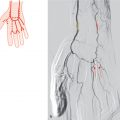
Fig. 13.3 The celiac trunk has four main branches (tetrapus), the fourth being an additional posterior pancreatic artery (10%). Schematic (a) and sagittal MIP CT (b). 1 Celiac trunk; 2 superior mesenteric artery; 3 posterior pancreatic artery.
13.2 Incomplete Celiac Trunk (9%)

Fig. 13.4 Hepatosplenic trunk; the left gastric artery is a separate branch of the aorta (5%). Schematic (a), axial MIP CT at the level of the celiac trunk (b), and coronal VR CT (c). 1 Left gastric artery; 2 hepatosplenic trunk; 3 splenic artery; 4 common hepatic artery.

Fig. 13.5 Gastrosplenic trunk; the common hepatic artery is absent or an independent branch of the aorta (3%). Schematic (a) and 3D VR CT image (b). The CT image shows the celiac trunk with the common hepatic artery as an independent branch of the aorta. 1 Left gastric artery; 2 splenic artery; 3 left renal artery; 4 superior mesenteric artery; 5 right renal artery; 6 common hepatic artery.
Related posts:


Stay updated, free articles. Join our Telegram channel

Full access? Get Clinical Tree


