17 Gastric Arteries
K.I. Ringe, S. Meyer
Textbooks usually list six arteries for the stomach and outline them in figures. The left and right gastric arteries anastomose along the lesser curvature. The left and right gastroepiploic arteries are found along the greater curvature. The fundus of the stomach is supplied by the short gastric arteries, which vary in number, and the pyloric region, by the gastroduodenal artery. The arterial anastomoses at the cardioesophageal (see esophageal arteries, Chapter 5) and at the gastroduodenal end of the stomach are extremely variable. In most cases, the left gastric artery is more prominent than the right.
17.1 “Normal” Blood Supply of the Stomach
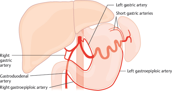
Fig. 17.1 “Normal” blood supply of the stomach. Schematic.
17.2 Right Gastric Artery
At the greater curvature, the right gastroepiploic artery is nearly always larger than the left, and the right gastroepiploic artery usually ends beyond the midline of the lower border.1–14
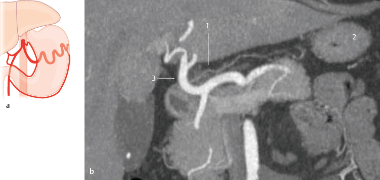
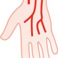
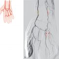
Fig. 17.2 “Normal” situation, origin from the common hepatic artery (~50%). Schematic (a) and coronal MIP CT (b). 1 Right gastric artery; 2 stomach; 3 common hepatic artery.
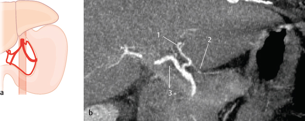
Fig. 17.3 Origin from the left hepatic artery (15%). Schematic (a) and coronal MIP CT (b). 1 Left hepatic artery; 2 right gastric artery; 3 right hepatic artery.

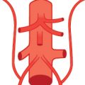
Fig. 17.4 Origin from the right hepatic artery (5%). Schematic.

Fig. 17.5 Origin from a middle hepatic artery or at the bifurcation of the proper hepatic artery (20%). Schematic (a) and CT images (b,c). Coronal VR (b) and MIP (c); the right gastric artery originates from the bifurcation of the proper hepatic artery. 1 Right gastric artery; 2 stomach.
17.3 Left Gastric Artery
The left gastric artery arises in approximately 90% of all cases from the celiac trunk and is usually the first branch. It is of great importance that in about one of four patients the left gastric artery participates in the blood supply of the liver, and in half of these cases it is an accessory hepatic but in the other half (11.5%) a replaced hepatic artery. The left gastric artery usually divides into two main branches, anterior and posterior.1,3,4,7,15,16 The right gastric artery anastomoses in most cases with the posterior branch of the left gastric artery.1,3,7,17

Fig. 17.6 “Normal” origin from the celiac trunk (~90%). Schematic (a), coronal VR CT (b), coronal MIP CT (c), and sagittal MIP (d). 1 Left gastric artery; 2 stomach; 3 celiac trunk.
Related posts:


Stay updated, free articles. Join our Telegram channel

Full access? Get Clinical Tree


