25 Obturator Artery
K.I. Ringe
The number of its variations increases considerably if all the different types of branching of the internal iliac artery are considered.1–7 To avoid confusion, the most common type, that is, the division of the internal iliac artery into two stems (Chapter 23), is taken as a basis for these figures. The percentages for the different types are averages. Accessory obturator arteries are not outlined. Small accessory branches or different origins with a trunk formation are very common. In such cases, arteries can cross the obturator nerve,7 which can be of relevance during pelvic lymph node dissection. The obturator artery supplies the muscles of the inner wall of the pelvis (obturator internus, psoas muscle) and has a pubic branch to the symphysis, which anastomoses with the obturator branch of the inferior epigastric artery. In Fig. 25.8, this anastomosis is very prominent and in Fig. 25.7 the inferior epigastric artery alone feeds the obturator artery. A major branch between the inferior epigastric artery and the obturator artery was formerly called the “arteria corona mortis.” This artery was sometimes lacerated during herniotomies, and because such arterial bleeding was difficult to staunch in former times, it could prove fatal. If it is not recognized in pelvic fractures, it may result in untreated arterial bleeding.8 After passing the obturator canal, the obturator artery divides into the anterior branch to the adductor muscles and the posterior branch to the hip joint.9
25.1 Origin from the Internal Iliac Artery (75%)
A small acetabular branch reaches the head of the femur through the ligament of the head of the femur. Normally this branch is not able to supply sufficient blood to the head of the femur in cases of an epiphysiolysis. Racial differences have been discovered in connection with the origin of the obturator artery from the external iliac artery. The data given here are derived from studies on North Americans and Europeans; however, in Japan the frequency of this anomaly seems to be halved.1 In females, this anomaly is assumed to be more frequent. The obturator artery can also originate from the femoral artery; it then turns backward through the lacuna vasorum into the pelvis.10 The anastomoses between the obturator artery and the inferior epigastric and other arteries of the leg are sometimes of importance when the external iliac artery is occluded in atherosclerosis.

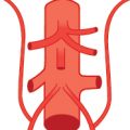
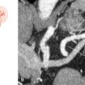
Fig. 25.1 The obturator artery branches from the stem of the internal iliac artery (15%). Schematic (a) and coronal oblique 3D VR CT (b). The CT shows the obturator artery branching from the stem of the internal iliac artery on the right side. 1 Obturator artery; 2 internal iliac artery.

Fig. 25.2 The obturator artery branches from the anterior stem of the internal iliac artery (25%). Schematic (a) and coronal oblique 3D VR CT (b). The CT shows the obturator artery branching from the anterior stem of the internal iliac artery on the right side. 1 Anterior stem; 2 obturator artery.
Related posts:


Stay updated, free articles. Join our Telegram channel

Full access? Get Clinical Tree


