Arteriovascular Disease and Ischemia
Spinal Dural Arteriovenous Fistulas
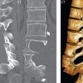
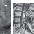
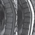
The type I spinal dural arteriovenous (AV) fistula is the most common vascular malformation of the spine, presenting mostly in older men in the thoracolumbar region. It is a direct shunt between the arterial and venous system (between a radiculomeningeal artery and a radicular vein), with drainage retrograde into perimedullary vessels (those surrounding the cord), leading to venous congestion of the cord. The arterial stasis and venous congestion cause chronic hypoxia and (typically) slowly progressive myelopathy from venous hypertension (Foix Alajouanine syndrome). Without therapy, the result can be irreversible paraplegia.
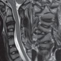
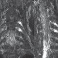
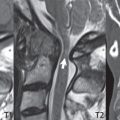
On MR, cord edema (with hyperintensity on T2-weighted scans, predominantly centrally in the gray matter—and thus H-shaped on axial images) and dilated perimedullary vessels are seen ( Fig. 3.112 ). The latter can be seen as numerous flow voids within the CSF adjacent to the cord on T2-weighted scans, and with enhancement post-contrast. Contrast enhancement of the cord itself may also be present. Contrast-enhanced MR angiography (MRA) can be used to directly visualize the tortuous, abnormally coiled, dilated perimedullary veins within the subarachnoid space ( Fig. 3.113 ).


Related posts:
Stay updated, free articles. Join our Telegram channel

Full access? Get Clinical Tree


