Infection and Inflammation


10.1055/b-0034-102689

Infection and Inflammation


Disk Space Infection


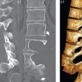
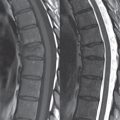
Disk space infection can be hematogenous in origin, or a complication of spine surgery. In the latter, the clinical presentation is usually several weeks following surgery, with delays in diagnosis common. Focal pain is a prominent clinical feature, but may be absent. In hematogenous seeding, it is stated that the disk serves as the initial site of infection in children, as it is richly vascularized, whereas in adults the initial site of infection is the subchondral bone or adjacent soft tissue. On CT, only late changes are noted, including disk space narrowing, cortical endplate destruction and irregularity, and an accompanying paraspinal soft tissue mass. On MR, the most common presentation of a disk space infection is that of a narrowed irregular disk ( Fig. 3.116 ), with focal high signal intensity on the T2-weighted scan (infected fluid pockets), enhancement of the disk space (other than the fluid), and a horizontal band of edema within the vertebral body both above and below (osteomyelitis), paralleling the infected disk space. The edema within the adjacent vertebral bodies will display enhancement on post-contrast scans obtained with fat saturation ( Fig. 3.117 ).

Fig. 3.116 Disk space infection with osteomyelitis, lumbar spine. On a sagittal T1-weighted midline image, abnormal low signal intensity is seen involving the vertebral endplates at the L2–3 level, with irregularity of the inferior L2 endplate, and poor visualization of portions of the superior L3 endplate. Abnormal prevertebral soft tissue is also present at this level (white arrow). On the corresponding sagittal STIR image, the disk space is noted to be narrowed, irregular, and with abnormal high signal intensity (small black arrows), the latter reflecting fluid within the disk space. The findings involving the disk space itself are consistent with infection, which is accompanied by abnormal signal intensity in the adjacent vertebral endplates (low on T1, high on STIR), consistent with osteomyelitis.
Fig. 3.117 Disk space infection with accompanying osteomyelitis (spondylodiscitis), extensive disease involvement. On sagittal images, there is fluid in the L3–4 disk space, seen as high signal intensity on the T2-weighted scan and by the lack of enhancement post-contrast. There is extensive edema (with accompanying abnormal contrast enhancement) throughout L3 and L4. These are characteristic findings in an advanced case of disk space infection, with the abnormality in the marrow indicative of osteomyelitis. There is pre-/paravertebral abnormal soft tissue (arrow), representing further disease extent, and loss of height of L3. There is also extension to L5 (thus involvement of 3 vertebral bodies, which is unusual), with abnormal marrow signal intensity and enhancement superiorly in L5 and involvement of the disk at L4–5 (with abnormal enhancement). The etiology in this instance was methicillinresistant Staphylococcus aureus (MRSA), accounting for the unusual extent of disease.

In early disease, the extent of edema within the adjacent vertebral bodies may be mild (or absent with discitis only), and in severe disease both the vertebral body above and below can be involved in their entirety. Although often less evident than on CT, the vertebral endplates will appear indistinct on MR in a disk space infection with accompanying osteomyelitis. A paraspinal soft tissue mass is also commonly present, of varying size, with enhancement post-contrast ( Fig. 3.118 ).

Fig. 3.118 Disk space infection, with extensive spread of infection into the adjacent soft tissues and epidural space. Abnormal enhancement, consistent with osteomyelitis, is seen throughout the L4 and L5 vertebral bodies. The L4–5 disk space is irregular and does not enhance, consistent with an infected fluid collection. There is extensive abnormal, enhancing prevertebral and paravertebral soft tissue, with extension to the epidural space and canal compromise on that basis.

Often CT-guided biopsy is performed in order to have material for bacterial culture, prior to starting antibiotic treatment. Once treatment with intravenous antibiotics is initiated, the changes on MR (reflecting recovery) lag behind that of the clinical course. Other causes of paraspinal infection, without accompanying discitis and osteomyelitis, include hematogenous seeding (e.g., with a psoas abscess) and following surgery, with infection in the operative bed posteriorly.


Tuberculous Spondylitis


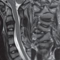
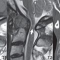
Tuberculous spondylitis follows a more indolent clinical course than pyogenic infection. In the United States this disease is seen primarily in immigrants (from Southeast Asia and South America) and in immunocompromised patients. The disease is primarily one of adults, and occurs due to hematogenous seeding. Differentiating features on imaging from pyogenic infection include inoculum in the anterior vertebral bodies, relative sparing of the disk space due to absence of proteolytic enzymes (early in the disease process) ( Fig. 3.119 ), involvement of multiple contiguous vertebral bodies (three or more levels in half of patients), and a large associated paraspinal mass. Although skip lesions are reported, these are uncommon (< 5%). Spread of disease occurs under the anterior longitudinal ligament. In long standing disease there will be extensive bone destruction.

Fig. 3.119 Spinal tuberculosis. Illustrated on sagittal images is a major distinguishing feature of tuberculous spondylitis, relative sparing of the disk space, despite involvement of the adjacent vertebral bodies. Note the prominent involvement of L4 and L5, with extension of disease to the superior/anterior portion of S1, all well visualized pre-contrast with abnormal low signal intensity, and post-contrast with abnormal enhancement. Although there is loss of disk space height, anteriorly the L4–5 disk is spared, which would not be seen in pyogenic disk space infection (the primary differential in this instance).


Epidural Abscess


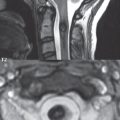
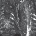
In recent years, epidural abscesses in the spine have become more frequent, with a subdural abscess, though, very uncommon. Infection can occur by hematogenous spread, direct extension (e.g., anteriorly from a disk space infection with osteomyelitis), or penetrating trauma. There may be associated meningitis and myelitis. MR is the modality of choice for detection and delineation, with CT of little value. Thickened inflamed (enhancing) soft tissue (phlegmon) is seen initially, progressing to a frank abscess (with fluid centrally). Contrast enhancement on MR can thus be homogeneous, or rimlike with central low signal intensity (pus). There can be canal compromise and cord compression, due to inflammation, granulation tissue, and any associated fluid collection ( Fig. 3.120 ). Lesions usually extend over several levels, and are often very extensive ( Fig. 3.121 ).

Fig. 3.120 Epidural abscess, cervical spine. An extensive infected epidural fluid collection is present, largest in bulk anteriorly in the high cervical region (*), but present both anterior and posterior to the cord, effacing the thecal sac. On the sagittal T2-weighted scan, this collection is slightly lower in signal intensity in comparison to CSF, and on the sagittal T1-weighted scan slightly hyperintense. The prominent peripheral enhancement (in part representing the displaced dura) is characteristic.
Fig. 3.121 Epidural abscess, thoracic spine. On the sagittal T2-weighted FSE scan, a posterior epidural mass with mixed signal intensity is noted to compress the cord anteriorly. The lesion is well delineated post-contrast with fat saturation, consisting of an extensive (infected) fluid collection outlined by a prominent enhancing inflammatory tissue margin.

Both sagittal and axial imaging are important, the former for definition of extent of disease, and the latter for improved assessment of the thecal sac and cord compression. Diffusion weighted scans (an abscess will demonstrate restricted diffusion), relatively recently available with reasonable image quality for spine MR imaging, are an important complementary imaging sequence ( Fig. 3.122 ).

Fig. 3.122 Epidural abscess, diffusion weighted imaging. An epidural fluid collection is identified, immediately posterior and to the left of the thecal sac in the thoracic region. There is marked compression of the thecal sac and mild cord deformity. The hyperintensity of the fluid collection on DWI is consistent with an abscess, which was proven pathologically. Restricted diffusion was confirmed on the ADC map (not presented). Post-contrast there is peripheral enhancement.

Only gold members can continue reading. Log In or Register to continue

Stay updated, free articles. Join our Telegram channel

Jun 14, 2020 | Posted by in NEUROLOGICAL IMAGING | Comments Off on Infection and Inflammation

Full access? Get Clinical Tree

Get Clinical Tree app for offline access