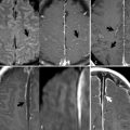
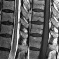
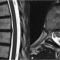
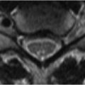
10 Arteriovenous Malformations Arteriovenous malformations (AVMs) are the most common cerebrovascular malformation, consisting of direct communication between the arterial and venous circulations without intervening capillaries. Their most serious complication is hemorrhage (4% annual rate) with a yearly mortality of 1%. Symptoms of AVMs include headache, seizures, and neurologic defects. These may be caused by mass effect or a steal phenomenon whereby blood flow is diverted from the surrounding parenchyma to an AVM, underperfusing the former. AVMs are associated with a variety of inherited conditions such as Sturge-Weber (port-wine stain, mental retardation, glaucoma, and seizures), Osler-Weber-Rendu (mucocutaneous telangiectasias and AVMs), and Wyburn-Mason (midbrain AVM, facial nevus in distribution of the trigeminal nerve, and retinal angioma ipsilateral to the facial nevus). The most common major feeding vessel is the middle cerebral artery, and 80% of lesions are supratentorial. Classically, an AVM is visualized on MRI as a tangled nidus (Fig. 10.1A, arrow) of dilated vessels supplied by enlarged feeding arteries, with multiple enlarged, tortuous draining veins. AVM arterial supply is most frequently pial but may be dural, especially in infratentorial malformations. Feeding arteries are identified by location and dilatation. As shown in Fig. 10.1B where the anterior cerebral artery is feeding an AVM, TOF MRA may be useful in localizing the feeding artery. Aneurysms within feeding arteries occur in ~10% of patients and often regress after AVM treatment. Draining veins (Fig. 10.1C,D black arrow
![]()
Stay updated, free articles. Join our Telegram channel

Full access? Get Clinical Tree


