Arthrogram, Shoulder
Spinal needle, 20- or 22-gauge, 3.5-in. length (approximately)
Lidocaine 1%.
Syringes, 10 and 20 mL, with short extension tubing.
Saline, sterile nonbacteriostatic.
Nonionic contrast, 20 mL (approximately), Omnipaque 300 or 360 or equivalent.
Indications.
Suspected rotator cuff tear, adhesive capsulitis, loose bodies. Injection of magnetic resonance (MR) contrast for MR arthrography.
Consent.
Risk of procedure is mainly due to possible allergy to contrast or local anesthetic. Bleeding and introducing infection are remote possibilities.
Scout Films.
Anteroposterior (AP) films of shoulder in internal and external rotation; axillary view.
Patient Position.
Patient lies supine with injured shoulder toward operator, arm supinated and palm up (externally rotated). Slight elevation of the shoulder with a pillow may help to rotate the glenoid labrum out of needle’s path to joint space.
Landmarks.
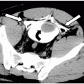
Begin at skin 1 cm inferior and 1 cm lateral to coracoid process. Aiming for the center of the joint, mark exact skin position with a radiopaque marker using fluoroscopy (Fig. 55-1). Under fluoroscopy, the glenoid should be facing slightly anteriorly and not seen in tangent; otherwise, the glenoid labrum will be in the way.
Preparation.
Make a skin indentation or indelible mark at desired puncture site. Prepare and drape the area using sterile technique. Administer local anesthetic.
Insert Needle.
Related posts:
Stay updated, free articles. Join our Telegram channel

Full access? Get Clinical Tree


