Myocardial Ischemia and Infarction
Jens Vogel-Claussen
Oleg M. Teytelboym
Etiology and Epidemiology.
Coronary artery disease is currently the number one cause of death of both men and women in the United States. Risk factors include smoking, diabetes, hypertension, dyslipidemia, and family history. It is caused by formation of atherosclerotic plaques within the walls of coronary arteries, which may impede vascular flow. Myocardial ischemia, like all tissue ischemia, results from excessive demand or inadequate supply of oxygen, glucose, and free fatty acids due to coronary artery luminal narrowing. Hemodynamically significant stenosis is present if there is a more than 50% luminal diameter narrowing of left main artery or a more than 70% narrowing of other vessels. As atherosclerotic plaque evolves, production of macrophage proteases and neutrophil elastases within the plaque can cause thinning of the fibromuscular cap that covers the lipid core. Increasing plaque instability coupled with blood-flow shear and circumferential wall stress can lead to plaque fissuring or rupture, especially at the junction of the cap and the vessel wall. Acute myocardial infarction results from a rupture of an unstable plaque and secondary thrombus formation which leads to occlusion of the lumen, most commonly occurring in the proximal portions of the coronary arteries. Unstable angina is characterized by rapid thrombolysis of this thrombus, which restores myocardial perfusion and prevents myocardial tissue death. Stable angina occurs due to significant luminal stenosis without plaque rupture and thrombosis.
Symptoms and Signs.
Chest pain is the cardinal symptom of myocardial ischemia. The pain commonly radiates to the left arm or the jaw. Patients may also complain of dyspnea, nausea, diaphoresis, and chest pressure. However, particularly in patients with diabetes, myocardial ischemia may be asymptomatic. Stable angina manifests as increased myocardial demand due to exercise or emotional stress, and is relieved by rest or sublingual nitroglycerin. Unstable angina is characterized by increasing frequency and severity of chest pain (crescendo-angina), chest pain at rest, and increasing need of antiangina medication occurring in patients with a history of stable angina. Patients with unstable angina have a 20% risk of acute myocardial infarction. Acute myocardial infarction is characterized by sudden onset (typically occurring over 30 minutes) of intense chest pain and pressure. Electrocardiography (ECG) is the cornerstone of rapid diagnosis. Elevation of myocardial enzymes is used to confirm the diagnosis.
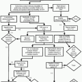
Clinical history, ECG changes, and cardiac enzyme analysis are sufficient for establishing the diagnosis of acute myocardial infarction in most cases. Imaging is primarily used to diagnose and evaluate stable and unstable angina in order to detect hemodynamically significant stenosis or to evaluate myocardial viability. Myocardial viability imaging can be used to distinguish scar from hypoperfused hibernating myocardium, in order to predict outcomes of revascularization.
Stress ECG has an approximately 80% sensitivity of detecting a critical coronary narrowing if the age-related target heart rate is obtained. Stress echocardiography has a
sensitivity of approximately 90% in detecting wall motion abnormalities. However, this technique is operator dependent and has a limited field of view. Plain chest radiographs are quite insensitive in detecting coronary pathology. In patients with severe disease, heavy coronary artery calcifications may show up as “railroad tracks” on chest radiographs projecting over the heart silhouette. Also ventricular enlargement, cardiac aneurisms, pleural effusions, and pulmonary edema as sequela from myocardial infarction can be evaluated with chest radiographs.
sensitivity of approximately 90% in detecting wall motion abnormalities. However, this technique is operator dependent and has a limited field of view. Plain chest radiographs are quite insensitive in detecting coronary pathology. In patients with severe disease, heavy coronary artery calcifications may show up as “railroad tracks” on chest radiographs projecting over the heart silhouette. Also ventricular enlargement, cardiac aneurisms, pleural effusions, and pulmonary edema as sequela from myocardial infarction can be evaluated with chest radiographs.
In this chapter we focus on computed tomography (CT), magnetic resonance imaging (MRI), conventional coronary angiography, and nuclear medicine imaging techniques for detection of ischemic heart disease.
Indications.
Rapid evolution of CT technology has brought cardiac CT to the forefront of cardiac imaging. Calcium scoring can be used for coronary heart disease screening and also typically performed as part of coronary computed tomographic angiography (CTA). CTA is excellent at accurately assessing the atherosclerotic burden and luminal narrowing, and has been shown to be superior to conventional coronary angiography in detection of nonobstructing plaque. Although the guidelines are rapidly evolving, currently coronary CTA is considered in patients with equivocal stress tests. Recently, however, some emergency rooms have started to use coronary CTA as a tool for rapid triage and management of patients with chest pain.
Protocol.
A multislice CT scanner and heart rate control are the necessary prerequisites for performing an adequate cardiac CT. A 16-slice CT allows an adequate examination in 60-70% of patients, particularly if the heart rate can be maintained in the low 60s. A 64-slice CT achieves adequate results in more than 80-90% of patients, even if the heart rate is in the 70s. The new generation of dual source CT scanners should permit adequate cardiac CT with heart rates up to 100.
Heart rate control can be achieved with β-blockers: 50-100 mg of oral metoprolol 45-90 minutes before the scan or 5 mg metoprolol IV boluses, every 5 minutes up to 20 mg, with the patient on the CT table. Contraindications to β-blockers include second-degree Mobitz or third-degree arteriovenous (AV) block and severe chronic obstructive pulmonary disease (COPD).
Calcium scoring is obtained by performing a breath-hold noncontrast gated coronary CT, and is based on the Agatston scoring algorithm which was initially developed for electron beam CT. CT threshold density of 130 HU is used to detect coronary calcification in each of the four main coronary branches (left main, left anterior descending, circumflex, and right coronary arteries). The score is computer generated by measuring the volume of coronary calcification and multiplying it by a factor (between 1 and 4) based on the peak attenuation value of the lesion.
Coronary CT angiography is obtained by performing a breath-hold gated CT after injection of 80 mL of iodinated contrast bolus, injected at 4 mL/second, followed by 40 mL saline chaser (this requires a dual head injector) to reduce high-density artifacts in the superior vena cava and right heart (mixing 5 mL of contrast with the saline bolus can significantly improve septal visualization, without significant artifact). The image acquisition delay for each patient is usually determined by live bolus tracking in the descending aorta and triggering the scan when contrast density is 180 HU. Alternatively, a small test bolus can be used to determine peak contrast density time and adding 4- 6 seconds to allow for larger volume of the actual bolus.
High radiation dose is the main drawback of the current 64-slice techniques (˜9.8-16.3 mSv for males and 13.5-22.6 mSv for females versus 3-10 mSv for cardiac catheterization). Radiation dose can be reduced by using prospective ECG gating in order to scan only during diastole; however, this technique may significantly degrade examination quality in patients with even mildly irregular heart rate. Dose modulation by reducing the dose during the systole can decrease the total radiation dose by 40%.
|