Intracranial Venous Thrombosis
Bradley R. Foerster
Myris Petrou
Cerebral venous sinus thrombosis (CVST) is an uncommon condition with serious potential clinical consequences especially if the diagnosis is delayed. CVST has a 5-15% mortality rate and generally occurs in young or middle aged adults, accounting for 1-2% of strokes in these patients. Risk factors include pregnancy, oral contraceptive use, dehydration, adjacent infection (sinuses or mastoids), coagulopathy, malignancy and trauma. In 20% of patients, no underlying etiology is found. CVST is most common in the superior sagittal sinus, followed in decreasing frequency by the transverse and sigmoid sinuses, cerebral veins, straight sinus, and cerebellar veins. Septic cerebral venous thrombosis is relatively rare in developed countries and should be aggressively treated with antibiotics.
Symptoms and Signs.
Unfortunately, presentation of cerebral venous thrombosis is highly variable depending on the location and burden of the thrombosis and presence of venous collaterals. Headache is the most common, and sometimes the only, symptom occurring in up to 95% of cases and usually precedes the onset of other neurologic deficits by days or even months. Focal neurologic deficits are the next most common sign and include central motor and sensory deficits, seizures, aphasia, and hemianopia. These deficits may fluctuate and can alternate. Patients presenting with the syndrome of pseudotumor cerebri (headache, vomiting, visual changes, and papilledema) or unexplained hemorrhagic infarction (bilateral or not follow an arterial vascular distribution) must be evaluated for cerebral venous thrombosis.
Septic cavernous sinus thrombosis has a distinct clinical picture with chemosis, exopthalmos, painful opthalmoplegia, third, fourth, and sixth nerve palsies. Complications include occlusion of the internal carotid artery and extension into dural sinuses or brain parenchyma. Because of its often rapidly fatal course, septic cavernous sinus thrombosis should be treated presumptively before imaging.
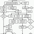
Differential Diagnosis.
Because of the highly variable presentation differential diagnosis includes arterial stroke, cerebral tumors, brain abscess, arteriovenous malformation, encephalitis, cerebral vasculitis, and seizure disorders.
Complications
Infarction and hemorrhage occur in approximately 35-50% of patients. The venous thrombosis increases venous and capillary pressure, resulting in decreased blood flow to the brain parenchyma. Breakdown of the blood-brain barrier and diapedesis of erythrocytes are thought to contribute to the hemorrhagic infarct.
Seizures can be focal or generalized and seen in 35-50% of patients and up to 75% in peripartum females.
Obstructing hydrocephalus can occur with cerebellar vein thrombosis, causing cranial nerve palsies and life-threatening hydrocephalus.
Encephalopathy and coma are seen in patients with extensive thrombosis or involvement of the deep venous system with bilateral thalamic involvement. Severe alteration of consciousness is the strongest predictor of poor clinical outcome.
Indications.
A noncontrast head computed tomography (CT) should be performed as the initial study for evaluation of patients presenting with acute neurologic symptoms. Unfortunately, noncontrast head CT is neither sensitive nor specific for the diagnosis of CVST but can help to exclude other life-threatening diseases. Computed tomographic venography (CTV) is a rapid and widely available modality for direct visualization of the intracerebral venous system to diagnose thrombosis. It is equal to magnetic resonance venography (MRV) for diagnosing CVST, but inferior to magnetic resonance imaging (MRI) in establishing other potential etiologies of the patient’s symptoms.
Protocol.
Routine noncontrast head CT should be performed first. CTV is performed after a short delay (˜60 seconds) following contrast administration with acquisition of thin slices for review on a three- dimensional (3D) workstation.
Related posts:
Stay updated, free articles. Join our Telegram channel

Full access? Get Clinical Tree


