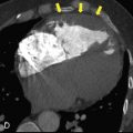
Cone-beam computed tomography (CBCT) has become one of the most dependable tools for assessing dental implants, surrounding bone, and important anatomical structures. However, titanium implants often create distortions in the image, commonly referred to as artifacts. These distortions can complicate interpretation and make it harder for clinicians to get a clear, accurate view of the implant site. Because of this, artifact reduction has become an essential part of modern dental imaging.
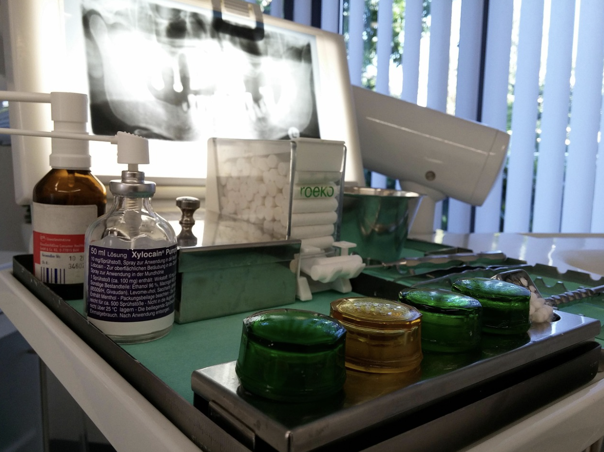
Image by peter-facebook on Pixabay
Many practices that focus heavily on implant care, including Rivertown Dental, emphasize mastering techniques that help minimize these distortions. Understanding how artifacts form—and how to reduce them—allows dental professionals to produce clearer scans, make more informed decisions, and provide better patient care. This article explains these methods in a simple and approachable way, while still offering meaningful clinical value.
Why Titanium Implants Create Imaging Challenges
Titanium is an excellent material for dental implants because it bonds well with bone and offers long-term durability. However, it also interacts strongly with X-ray beams. When the CBCT unit sends X-rays through titanium, some of these rays scatter, while others become overly absorbed, leading to streaking or distortion in the final scan.
This process, known as beam hardening, filters out lower-energy photons and leaves higher-energy photons dominating the beam. As a result, the detector receives inconsistent data, which appears as warped or unclear areas around the implant. Even though this is a normal physical effect, understanding it helps dentists anticipate and manage these imaging challenges.
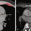
Common Types of Artifacts Seen Around Titanium Implants
Streak artifacts are among the most noticeable. They show up as thin bright or dark lines radiating from the implant. These streaks can hide important anatomical details or make bone margins harder to evaluate.
Another frequent issue is shadowing, where bone or soft tissue around the implant appears darker than expected. This can mimic bone loss or other complications, making interpretation more difficult. Recognizing these patterns helps clinicians determine which artifact reduction strategies to use.
Equipment-Based Strategies for Reducing Artifacts
Using advanced CBCT machines is one of the most effective ways to minimize artifacts. Newer units often include specialized hardware and software designed to handle metal interference. These improvements help produce more reliable images, even in difficult cases.
Enhanced detector sensitivity and improved beam filtration also play an important role. Modern detectors reduce noise, while filtration removes low-energy photons that contribute heavily to beam hardening. Together, these updates significantly reduce distortion around titanium implants.
Adjusting voxel size is another helpful strategy. Smaller voxels provide more detail but may increase noise, while larger voxels offer smoother images but less precision. Selecting a balanced voxel size creates clearer, more dependable results.
Advanced reconstruction tools, such as Metal Artifact Reduction (MAR) algorithms, digitally correct distortions caused by metal. These algorithms analyze the data and reduce streaking, producing a clearer and easier-to-interpret final scan.
Technique-Based Strategies During Image Acquisition
Proper patient positioning is essential to reducing artifacts. A straight, stable head position helps the X-ray beam pass through the implant in a predictable and consistent way. Even minor positioning errors can worsen distortion.
Adjusting exposure settings is another useful method. Increasing the kilovoltage (kV) allows the beam to pass more efficiently through titanium, reducing streaking. The goal is to improve clarity while still keeping radiation levels as low as possible.
Using a smaller field of view (FOV) helps limit unnecessary radiation and reduces the volume of scanned metal. Focusing the scan on the exact area of interest produces clearer, more specific images and reduces artifact intensity.
Software and Post-Processing Options
Metal Artifact Reduction (MAR) filters can significantly improve clarity by correcting distortions created by metal objects. They work by analyzing the data and attempting to reconstruct missing or distorted information. While helpful, these filters should be used moderately to avoid over-smoothing important details.
Modifying the slice thickness during viewing can also improve accuracy. Thicker slices often reduce visible streaks, while thinner slices reveal more detailed structures. Switching between the two helps balance clarity and precision.
Changing the viewing angles in the software gives multiple perspectives of the same region. Some angles naturally reveal fewer artifacts, making it easier to detect bone changes or evaluate implant integration.
Clinical Strategies for Improving Interpretation
Combining CBCT results with clinical observations gives a more complete picture of the patient’s condition. Measurements, visual exams, and symptoms help confirm whether an unusual shadow is a true finding or simply an artifact.
Using supplementary radiographs, such as periapical X-rays, adds additional layers of information. These images can help verify areas that appear distorted on CBCT scans and support more accurate diagnosis.
In more complex cases, consulting with a dental radiologist can be invaluable. Radiologists have specialized training in identifying and working around artifacts and can provide deeper insight into unclear or distorted images.
Practical Tips for Clinicians and Technicians
- Follow a consistent scanning protocol. When all team members use the same settings, positioning practices, and review steps, image quality becomes easier to control and maintain. Consistency also supports reliable comparisons between scans taken at different times.
- Keep imaging software updated. Many CBCT systems receive regular updates that improve reconstruction quality or enhance MAR features. Staying current with these updates ensures the equipment performs at its best.
- Invest in ongoing staff training. Clear, consistent technique from every technician improves the accuracy of scans. Even small mistakes—like slight positioning errors—can increase artifact severity, so continued training is essential.
Sharper Images, Smarter Decisions
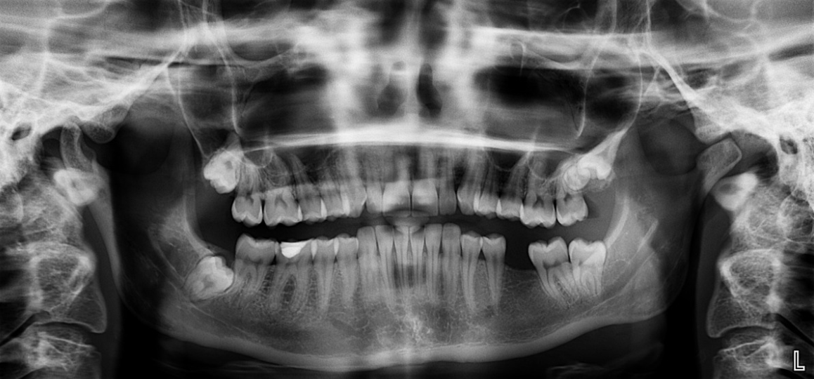
Image by youllneverknow on Pixabay
Reducing artifacts in CBCT imaging around titanium dental implants is more than a technical concern—it directly affects diagnostic accuracy and treatment success. By understanding how artifacts form and applying the right combination of equipment settings, patient positioning, and software tools, clinicians can create clearer, more reliable scans.
As CBCT technology continues to evolve, artifact reduction will only improve, giving dental professionals even greater confidence in their diagnostic decisions. With better images comes better planning, safer treatment, and improved outcomes for every patient who relies on implant care.
Related posts:


Stay updated, free articles. Join our Telegram channel

Full access? Get Clinical Tree


