Key Points
- ▪
The chest radiograph allows for determination of overall heart size assessment as well as of some degree of specific chamber size assessment.
- ▪
On the chest radiograph, the heart size is understood in the context of the chest size of the patient, affording some measure of indexing for body size.
- ▪
Limitations of absolute heart size assessment by the chest radiograph are numerous, but contingent on standard/comparable degrees of inspiration, serial assessment of heart size is useful.
Accurate assessment of heart size, and particularly of heart chamber size, requires the use of techniques such as echocardiography, computed tomography, or magnetic resonance imaging. However, estimates of the overall heart size are still very useful clinically, because, as a general rule, “cardiomegaly is disease.”
Methods to Assess Cardiac Size
Methods for assessment include the following:
- □
Gestalt: By integrating cardiopericardial silhouette (CPS) frontal plane area, cardiac contour, and ancillary findings, an experienced and able reader can appreciate an abnormally large CPS, with an accuracy favorable to more time-consuming measurement techniques.
- □
Cardiothoracic ratio (CTR; equivalent to transverse cardiac diameter divided by greatest internal chest width) ( Graphic 4-1 ): Several numbers are used as benchmarks to determine enlargement of the CPS: 0.50, 0.55, and 0.60. Obviously, a lower cutoff value is more sensitive but less specific. Generally, 0.50 is accepted as the upper limit of normal CTR. CTR correlates poorly with height, fairly with body weight, and best with height and weight together. There is a surprisingly wide range of normal values. Chest width is not a very useful index of body build, and its use introduces another noncardiac potential error; therefore, transverse cardiac dimension alone may be a better measure. Tables are available in some radiology texts.
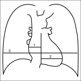
Graphic 4-1
Cardiothoracic ratio: maximal cardiac transverse diameter (1 + 2) divided by internal diameter of the chest cage (3).
- □
Frontal plane area: This has the best correlation (vs. gestalt and CTR) with cardiac volume but is time-consuming and seldom used.
- □
Cardiac volume: Currently cardiac volume is assessed by echocardiography, cardiac magnetic resonance, cardiac computed tomography, or contrast ventriculography, and it is almost never quantitatively assessed from a chest radiograph. However, validated formulas are available that calculate cardiac volume from measurements of cardiac dimension along its long axis, transverse axis (posteroanterior radiograph), and depth (lateral radiograph). In routine clinical practice, they are no longer used.
Related posts:


Stay updated, free articles. Join our Telegram channel

Full access? Get Clinical Tree


