Chapter 36 Atlantooccipital Joint Intraarticular Injection
Note: Please see page ii for a list of anatomical terms/abbreviations used in this book.
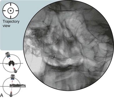
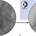
 Trajectory View (Figure 36–1)
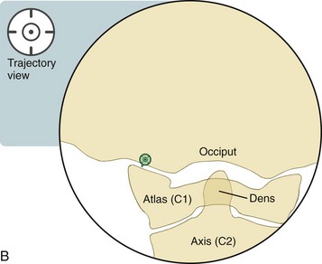
 Notes on Positioning in the Trajectory View


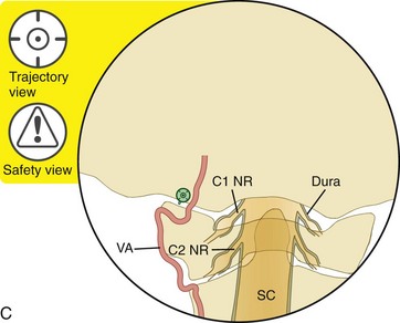
 Trajectory View Safety Considerations


Place the patient in the prone position with the neck slightly flexed and the head supported.
Confirm the level (with the anteroposterior view).
Oblique the fluoroscope 25 to 30 degrees ipsilaterally (to the left, in this case).
Related posts:
 Atlantoaxial Joint Intraarticular Injection
 Lumbar Zygapophysial Joint Nerve (Medial Branch) Radiofrequency Neurotomy, Posterior Approach
Stay updated, free articles. Join our Telegram channel

Full access? Get Clinical Tree


