- •
Location of defect on the atrial septum
- •
Relationship of the defect to the superior and inferior vena cava (sinus venosus type of atrial septal defect)
- •
Relationship of the defect to the atrioventricular valves (primum atrial septal defect)
- •
Direction of shunting across the defect
Anatomy and Anatomical Associations
The atrial septum, the part of the heart that separates the left from the right atrium, is not formed as a single structure. It is made up of embryological components that fuse in fetal and early postnatal life to fully separate the left from the right atrium. With the exception of a patent foramen ovale (PFO), any residual communication across the atrial septum after birth is considered an atrial septal defect (ASD).
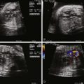
There are four subtypes of ASD, each named according to its embryological origin and the anatomical location on the atrial septum. A secundum ASD is a residual communication in the area of the fossa ovalis ( Figure 8-1 ). A primum ASD (see Chapter 9 ) is seen as part of the spectrum of disease associated with endocardial cushion defects or atrioventricular canal defects. Sinus venosus ASDs may be of the superior vena cava (SVC) or the inferior vena cava (IVC) type and are related to incomplete inclusion of the sinus venosus portion of the atrial septum. A coronary sinus ASD is located at the entrance of the coronary sinus into the right atrium and is synonymous with the finding of an unroofed coronary sinus.

The subtypes of ASD may be seen in isolation or as part of more complex congenital heart disease. A coronary sinus ASD may be associated with the presence of a left SVC.
Frequency, Genetics, and Development
ASDs are among the most common congenital heart defects, occurring at a rate of 1 in 1500 live births. Isolated secundum defects make up 6% to 10% of all congenital heart disease in the pediatric population. For an isolated ASD, the female-to-male ratio is approximately 2 : 1.
The development of the atrial septum occurs from the fourth to the eighth week of gestation. The septum primum, a thin membrane, grows from the roof of the primordial atrium toward the endocardial cushions. The ostium primum, the communication that exists between the encroaching septum primum and the endocardial cushions, closes as the septum primum fuses with the endocardial cushions. At the same time, programmed cell death occurs in the more posterior portion of the septum primum, creating the ostium secundum, which allows continued communication between the right and the left atrium. The septum secundum, an infolding of muscular tissue from the ventral wall of the primordial atrium, forms just to the right of the septum primum. By the eighth week of development, fusion of the septum primum and the septum secundum generally has occurred such that the only remaining communication through the septated atria is the foramen ovale, a flap valve formed by the attachment of the septum primum onto the septum secundum. The fossa ovalis is the region of the flap of the foramen ovale as seen from the right atrial side. As the septum primum and septum secundum form, a separate portion of the primordial atrium, the sinus venosus, splits into a right and a left horn. The right horn is the larger of the two and eventually forms the inflow of the IVC and SVC into the right atrium. The left horn fuses with the septum primum and forms the opening of the coronary sinus.
A secundum ASD is thought to arise from abnormal or excessive resorption of the septum primum during the period of programmed cell death. They can occur anywhere along the septum primum but typically occur in the region of the foramen ovale and fossa ovalis.
A sinus venosus ASD forms as a result of incomplete inclusion of the right horn of the sinus venosus into the right atrium and abnormal formation of the septum secundum. These defects are often associated with anomalous pulmonary venous connections and can have significant left-to-right shunting after birth. An ASD involving the coronary sinus results from incomplete fusion of the left horn of the sinus venosus with the septum primum.
An isolated ASD is not generally associated with syndromes or inheritable genetic anomalies, although there are some exceptions and some families with a inheritable form of ASD have been reported. One particular syndrome, Holt-Oram syndrome, is characterized by abnormalities of the upper extremities and cardiac septal defects, predominantly ventricular septal defects or secundum ASD. This syndrome is due to a mutation in the transcription factor TBX5. Other genetic associations include mutations in the genes NKX2-5 and GATA4. In addition, a missense mutation on chromosome 14q12 coding for myosin heavy chain has been shown to be responsible for a subset of inherited ASDs.
Fetal Physiology
The normal fetal circulation includes flow through the foramen ovale from the right atrium into the left atrium. Oxygen-rich blood returning from the placenta by way of the umbilical vein is preferentially directed by the eustachian valve across the atrial septum and into the left atrium. This blood then traverses the mitral valve and is ejected by the left ventricle, allowing the most highly oxygenated blood in the fetus to reach the most vital organs such as the developing fetal brain. The presence of an ASD does not influence this normal shunting in any way and is, therefore, of no hemodynamic consequence. In the absence of associated cardiac defects, an isolated ASD will not have a significant impact on fetal physiology.
Prenatal Management
Owing to the normal fetal flow of blood across the atrial septum, isolated ASDs are very difficult to diagnose in the fetus. However, the fetal circulation is such that an isolated ASD does not have an impact on fetal physiology and, thus, prenatal diagnosis is not essential.
Related posts:


Stay updated, free articles. Join our Telegram channel

Full access? Get Clinical Tree


