Chapter 20 Axial magnetic resonance imaging
Internal neural relations
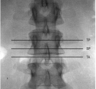
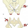
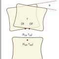
Axial magnetic resonance imaging (MRI) scans of the lumbar spine are typical taken through three standard locations at any given segment. The scans can be through the pedicles (transpedicular), below the pedicles (subpedicular) or through the intervertebral disc and zygapophysial joints (transarticular) (Fig. 20.1).
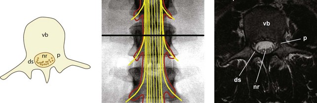
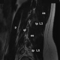
Each of these scans intersects slightly different elements contained within the vertebral canal. Transpedicular scans will intersect the dural sac and the nerve roots of the cauda equina (Fig. 20.2). In these scans, the pedicles will be evident projecting from the posterior surface of the vertebral body. These bones present as a grey signal. Posteriorly, various parts of the laminae and spinous process will complete the vertebral canal. Within the vertebral canal, the dural sac will appear as a black circle or oval, enclosing a white signal generated by the cerebrospinal fluid within the sac. Inside the sac, the nerve roots of the cauda equina appear as block dots, because the nerves appear as if they have been transected and looked at end-on. The nerve roots typically appear towards the posterior surface of the dural sac because patients typically lie supine when the scan is taken, and the nerve roots ‘fall’ to the rear.
Subpedicular scans will intersect the nerve roots of the cauda equina but also the spinal nerves of the segment, as they run through the intervertebral foramen of the segment (Fig. 20.3). The scan will show the vertebral body anteriorly, and the lamina on each side posteriorly. Behind the centre of the vertebral body, the dural sac will appear as a dark ring, with the nerve roots of the cauda equina appearing as black dots lying in a sea of white signal formed by the cerebrospinal fluid. Lateral to the dural sac a dark signal will appear, behind the vertebral body, created by the spinal nerve. Depending on exactly where the scan is taken, the spinal nerve may be accompanied by a sleeve of dura mater, which in turn may contain a sliver of cerebrospinal fluid surrounding the nerve.
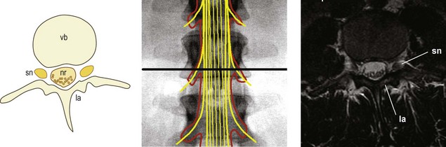
Transarticular scans will intersect the dural sac and cauda equina centrally, but will also show the spinal nerve or ventral ramus leaving the vertebral column and entering the psoas major muscle (Fig. 20.4
Stay updated, free articles. Join our Telegram channel

Full access? Get Clinical Tree


