32 Axillary Artery
B. Meyer, L. Sonnow
Enormous differences exist in the varieties of the axillary artery. Racial differences seem to play a role, but more important are the variations in the trunks’ names. The percentages given should be taken as rough estimates. The variations shown in Section 32.2 have an overall frequency of approximately 50%, which is lower than the sum of the subtypes shown in Fig. 32.2, Fig. 32.3, Fig 32.4, and Fig. 32.5 because these subtypes may occur in combination (Fig. 32.6). More exact data are not available.1–15
32.1 Normal Type as Shown in Textbooks (<10%)
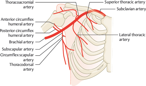
Fig. 32.1 Normal type as shown in textbooks (<10%). Schematic.
32.2 Trunk Formation of Some Branches (50%)

Fig. 32.2 The lateral thoracic artery forms a trunk with the thoracoacromial artery (~10%). Schematic (a) and VR CTA (b). 1 Axillary artery; 2 thoracoacromial artery; 3 common trunk; 4 lateral thoracic artery; 5 thoracodorsal artery; 6 circumflex scapular artery; 7 posterior circumflex humeral artery.

Fig. 32.3 The lateral thoracic artery originates with the subscapular artery (~10%). Schematic (a) and VR CTA (b). 1 Subscapular artery; 2 lateral thoracic artery; 3 circumflex scapular artery.

Fig. 32.4 The subscapular artery forms a trunk with the posterior circumflex artery of the humerus (~20%). Schematic (a) and VR CTA (b). 1 Posterior circumflex humeral artery; 2 lateral thoracic artery; 3 thoracodorsal artery; 4 circumflex scapular artery; 5 subscapular artery.

Fig. 32.5 Both circumflex arteries of the humerus form a trunk (20%). Schematic.

Fig. 32.6 Combination of the types shown in Fig. 32.2, Fig. 32.3, Fig. 32.4, and Fig. 32.5 (e.g., combination of Fig. 32.2 and Fig. 32.5). Schematic.

Fig. 32.7 The majority of branches originate as a common trunk from the axillary artery (2%). Schematic (a) and VR CTA (b). 1 Axillary artery; 2 common trunk; 3 lateral thoracic artery; 4 thoracodorsal artery; 5 circumflex scapular artery; 6 posterior circumflex humeral artery.
Related posts:


Stay updated, free articles. Join our Telegram channel

Full access? Get Clinical Tree


