Presentation and Presenting Images
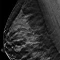
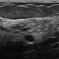
( ▶ Fig. 20.1, ▶ Fig. 20.2, ▶ Fig. 20.3, ▶ Fig. 20.4)
A 57-year-old female with a history of left ductal carcinoma in situ (DCIS) treated with segmentectomy and radiation therapy presents for routine screening mammography.
20.2 Key Images
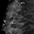
( ▶ Fig. 20.5, ▶ Fig. 20.6, ▶ Fig. 20.7)
20.2.1 Breast Tissue Density
The breasts are heterogeneously dense, which may obscure small masses.
20.2.2 Imaging Findings
The imaging of the right breast is normal (not shown). The left breast demonstrates postsurgical changes with architectural distortion and surgical clips (solid arrows in ▶ Fig. 20.6) in the left breast at the 2 to 3 o’clock position and a surgical clip (broken arrow in ▶ Fig. 20.6) in the left axilla. Fine radiodense particles project over the left axilla (circle in ▶ Fig. 20.6). The left breast tomosynthesis movie demonstrates that the fine densities are in skin folds of the left axilla consistent with deodorant artifact ( ▶ Fig. 20.7).
20.2.3 BI-RADS Classification and Action
Category 2: Benign
20.3 Differential Diagnosis
Deodorant artifact: The radiodense particles localize to the skin folds of the axilla. The quality of the radiodense particles also suggest a metallic quality; a metallic compound is a component of deodorant.
Skin calcifications: The radiodense particles appear on the surface of the skin and are not contained within the skin.
Axillary calcifications : The radiodense particles are on the surface of the skin and do not localize in the axilla, such as would be seen in nodal calcifications.
20.4 Essential Facts
Although the finding is not apparent on the left craniocaudal digital breast tomosynthesis (CC DBT) movie, it is well seen on the left mediolateral oblique (MLO) DBT movie. The finding corresponds directly with the lateral-most skin surface and the lateral-most MLO DBT slice (slice 1 of 81 in ▶ Fig. 20.7).
It is important for patients to thoroughly cleanse the skin of the breast and axilla prior to mammography to prevent artifacts.
Deodorant artifacts may be mistaken for axillary calcifications and may cause a patient to be recalled.
Prevention of this artifact is easily remedied by washing the axilla and if not done well, may be identified as an artifact with tomosynthesis imaging.
20.5 Management and Digital Breast Tomosynthesis Principles
Thorough preparation for conventional mammographic and tomosynthesis imaging involves the removal of deodorant and lotion, which may cause artifacts that mimic calcifications. In some cases, the artifacts mimic suspicious calcifications that may prompt a recall for biopsy.
Most radiology departments recommend that patients do not wear powder, deodorant, or lotion on the day of mammography.
Tomosynthesis imaging localizes deodorant to the skin folds rather than the skin. The location of the deodorant is not within the dermis of the axilla but on the skin surface.
Tomosynthesis evaluation of deodorant artifact can prevent unnecessary biopsies and reduce recall for this benign finding.
20.6 Further Reading
[1] Geiser WR. Artifacts in digital mammography. In: Whitman GJ, Haygood TM, eds. Digital Mammography: A Practical Approach. Cambridge, England: Cambridge University Press; 2012:80

Fig. 20.1 Left craniocaudal (LCC) mammogram.
Stay updated, free articles. Join our Telegram channel

Full access? Get Clinical Tree


