Some soft tissue structures are visible on a normal abdominal X-ray. Here the edges of the liver, kidneys and psoas muscles are clearly seen. Identifiable bones include the ribs, spine, pelvis and femora
16.2 Small bowel
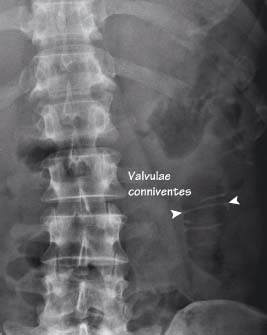
Normal small bowel is often more centrally placed than the colon. It can sometimes be clearly identified by the presence of visible valvulae conniventes (mucosal folds that pass across the whole width of the small bowel)
16.3 Large bowel
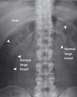
The large bowel (arrowheads) is often seen more peripherally than the central small bowel. The colonic wall is lined by haustra (mucosal folds that do not pass across the full width of the lumen)
16.4 Dense structures
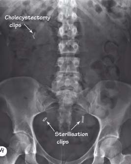
Calcified and other dense structures can often be seen on an abdominal X-ray. This patient has had two previous operations – a sterilisation procedure and a cholecystectomy. Clips left in place for these operations are clearly visible
Related posts:


Stay updated, free articles. Join our Telegram channel

Full access? Get Clinical Tree


