-“cow”, which can be milked every day) in the form of pertechnetate (TcO4–) which has a chemistry that is favorable for a number of coupling reactions. Thus 99mTc may be used coupled to phosphonate compounds for bone scintigraphy (Figure 51), to HIDA for biliary scintigraphy, to mercaptoacetyl-triglycin (MAG3) for renography, to albumin-aggregates for perfusion studies, for example lung perfusion; coupled to colloids for labeling of macrophages in liver, spleen, and bone marrow, and coupled to glucoheptonate or hexametazime for brain scintigraphy, to mention a few applications.
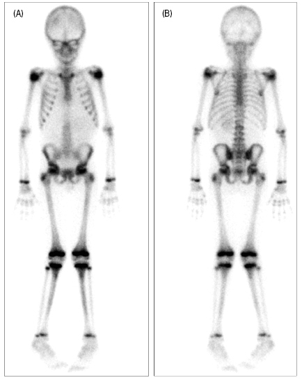
(A) is recorded with the boy’s front in contact with the gamma camera; (B) with the back and buttock in contact. Note the high signal intensity from growth plates and other sites of growth. By comparing the two images it is clearly seen that the recorded signal intensity is dependent on the distance from the camera.
The Gamma Camera
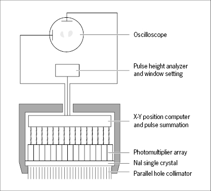
The basic design of a gamma camera, as used for diagnostic scintigraphic imaging, is shown in Figure 52. The γ-photon detector is a large single crystal of sodium iodide, doped with thallium. A collimator consisting of a lead plate with numerous closely spaced holes is mounted in front of the crystal. The holes may be parallel or they may be diverging towards the patient in order to obtain a larger field of view or converging to produce an enlarged image with more details. The collimator absorbs γ-photons which do not travel parallel or nearly parallel with the axis of the holes. Thus, the collimator defines, for each point in the crystal, the direction of incident γ-photons.
Stay updated, free articles. Join our Telegram channel

Full access? Get Clinical Tree


