2 Basic Physical and Technical Principles
You now know how to adjust the ultrasound machine, how to optimize the image quality, and how to use the transducer. You are ready to begin scanning.
If you like, you may skip the following section and return to it later if you need to. It provides a brief review of the basic physical and technical principles of diagnostic ultrasound.
LEARNING GOALS
 Understand the production, propagation, and detection of ultrasound.
 Learn the differences between A-mode, B-mode, and M-mode scanning.
 Be able to recognize and distinguish artifacts.
Ultrasound
Definitions
Sound is a mechanical wave that travels longitudinally through an elastic medium.
Ultrasound is sound at a frequency beyond the range of human hearing. The frequency range of diagnostic ultrasound is between 1 and 20 MHz.
Propagation of sound
Reflection and refraction
When sound encounters a boundary between two media of different densities, some of the sound “bounces” back toward the source as an echo. The angle of incidence of the sound is equal to the angle at which the echo is returned. This phenomenon is called reflection. The rest of the sound continues to travel through the second medium but is deflected from its original path. This phenomenon is called refraction.
K E Y P O I N T
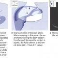
The spleen provides an acoustic window for scanning the left kidney.
Impedance
Acoustic impedance is the measure of resistance to the propagation of sound waves. It is the product of the density of a medium and the velocity of sound in the medium. When there is a large difference in acoustic impedance between two adjacent media, a large portion of the sound will be reflected at the interface between the media.
Absorption
As a sound wave propagates through matter, some of its energy is converted by friction into heat. This loss of sound energy is called absorption.
Scatter
Besides reflection and refraction, scatter is another phenomenon that is important in the propagation of ultrasound. When ultrasound waves encounter a nonhomogeneous medium or a “rough” surface, a small part of their energy is scattered away from the object, in random directions, while most of the sound continues to propagate. In diagnostic ultrasound, some of this scattered sound is radiated back toward the transducer and contributes to image formation.
Production and detection of ultrasound waves: the pulse-echo principle
Diagnostic ultrasound is based on the pulse-echo principle. The smallest functional unit of the ultrasound transducer is the piezoelectric crystal. This crystal has the property of converting electrical oscillations into mechanical vibrations, and vice versa. Thus, when the crystal is exposed to an alternating electric current, it will undergo mechanical deformation and generate sound waves. Conversely, when sound waves strike the crystal, they deform it and cause it to generate electrical impulses. One crystal can perform both functions in an alternating fashion.
K E Y P O I N T
The piezoelectric crystals in a transducer emit sound pulses and also receive the echoes.
First the piezoelectric crystal is exposed to an alternating electrical field, causing it to vibrate. The transducer emits a short, intense burst (pulse) of sound. Immediately thereafter, the transducer switches to the “listening” mode. The echoes reflected from different interfaces successively return to the crystal and cause it to vibrate. These vibrations are converted to electrical impulses, which are used to reconstruct an image.
Diagnostic ultrasound: propagation of ultrasound in biological tissue
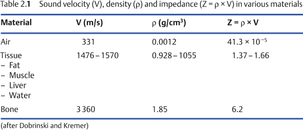
From an ultrasound standpoint, the human body is composed of three basic materials: gas, soft tissues, and bone (Table 2.1).

Stay updated, free articles. Join our Telegram channel

Full access? Get Clinical Tree


