Basics of SPECT Scanning: Emission and Transmission
Timothy G. Turkington
David R. Gilland
Single-photon emission computed tomography (SPECT) imaging provides cross-sectional images of the distributions of single-photon-emitting radiotracers. In all such tracers, one or more photons are emitted during the nuclear decay. The energy of the radiation is typically between 70 and 364 keV. The most commonly used radionuclide, technetium-99m (99mTc), emits a 140-keV γ-ray.
A gamma camera is used to acquire the data. This device uses a lead collimator (a flat slab with thousands of parallel holes), a sodium iodide (NaI) scintillator, and photomultipliers in a two-dimensional array covering the back of the scintillator. The collimator restricts the radiation so that only those photons emitted from the body in a particular direction are transmitted to the scintillator. The combination of NaI and photomultipliers, plus processing electronics, yields an energy value and coordinates (where the photon hit the detector) for each detected event. The accumulation of events over time yields a projection view of the radiotracer in the body.
For SPECT, multiple projection views are acquired all around the body. This requires that the detector (or detectors) be mounted on a rotatable gantry (see Fig. 4.5). Once views are obtained from a series of angles around the body, the data are reconstructed into multiple transaxial images. Each pixel in these images represents (with varying degrees of accuracy) the amount of radiotracer accumulated in the corresponding small region of the body. The multiple transaxial slices can be reformatted into coronal or sagittal views. Other oblique views are used, for example, to display the myocardium in perfusion studies.
SPECT images are limited by several physical factors. The first is the limited number of counts obtained during a scan. This leads to statistical noise in the reconstructed images. The second is the spatial resolution, limited by the shape of the holes in the collimator. The third is the occurrence of scatter events, such as when a photon scatters in the body and is subsequently detected, appearing to have originated at an incorrect location. Finally, attenuation occurs when the emitted photons are not detected because of scatter (single or multiple) or absorption. For most studies, attenuation is the largest obstacle to accuracy in SPECT images. Methods to correct attenuation of emitted photons in the body rely on transmission scanning. Several transmission-scanning methods have been implemented.
Introduction
Single-photon emission computed tomography (SPECT) imaging is based on the emission of single photons (γ-rays or x-rays) produced in the decay of certain radionuclides. The most commonly used radionuclide for SPECT imaging is technetium-99m (99mTc). The γ-ray emitted from 99mTc has an energy of 140 keV. A summary of some radionuclides used for SPECT is given in Table 4.1.
Table 4.1 Radionuclides Commonly Used in Spect Imaging | ||||||||||||||||||||||||||||
|---|---|---|---|---|---|---|---|---|---|---|---|---|---|---|---|---|---|---|---|---|---|---|---|---|---|---|---|---|
|
The data used to form SPECT images are acquired with a gamma camera. The detection of radiation by a gamma camera is depicted in Figure 4.1. A photon is emitted from a radiotracer molecule in a random direction and leaves the body. The gamma camera includes a lead collimator with multiple parallel holes.
These holes restrict the acceptance of photons so that only radiation emitted (roughly) perpendicular to the face of the gamma camera will be detected; the rest of the protons are stopped by the lead. The photons that make it through the collimator holes eventually hit the detector material, which is a sheet of thallium-doped sodium iodide [NaI(Tl)]. The vast majority of systems use 3/8-inch (9.5-mm) thick NaI(Tl), although thicker crystals are available and are more appropriate for energies greater than 140 keV. The radiation penetrates the crystal to a depth that depends on the energy of the radiation. The vast majority of 140-keV photons will stop within 3/8 of an inch, which justifies this thickness for 140 keV and lower energies. When a photon stops, some of its energy is released in the form of visible light. This light is measured by photomultipliers on the back of the crystal. The energy of the incident particle is accurately estimated from the sum of all the light energy measured in the photomultipliers. In addition, the position where the photon hit the detector is determined to 3.5- to 4.5-mm accuracy by an electronically implemented algorithm that compares the relative amount of light measured in each photomultiplier. The result for each incident photon that stops in the detector is a set of three numbers: two give the position of the photon in the scintillator (x and y), and the third gives its energy E.
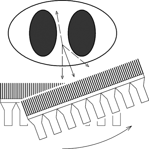 Figure 4.1 Detection of a photon by a gamma camera. Four different emission photon trajectories are depicted. Only one (indicated by the solid line) will be detected at this camera angle because of the lead collimator. |
As detected events accumulate, a projection view of the radiotracer in the body develops. The longer the acquisition, the more precisely this image represents the actual projection of the radiotracer distribution. SPECT imaging requires the acquisition of a series of these projections at successive angles around the body. To achieve this, the camera must be mounted on a gantry on which it can orbit the body. A complete data set requires an orbit of 180 degrees, and many acquisitions use 360 degrees of angular coverage. A typical angular increment is 3 degrees, which results in 60 total projections for a 180-degree orbit. Projection views and reconstructed slices from a cardiac SPECT study are shown in Figure 4.2. In this case, the field of view of the camera in the axial direction is 40 cm, much larger than necessary to image the heart.
Compared with positron emission tomography (PET), SPECT acquisition is a less efficient way of acquiring data, because only the photons leaving the body (roughly) perpendicular to the detector (about 1/1000, depending on the specific collimator design) will pass through the collimator and be detected. With PET, any photon pairs leaving the body in the same plane as the detector ring will be detected. Another disadvantage of SPECT is that only a single projection view is acquired at a time, whereas in PET all angles are acquired simultaneously.
Most single-photon emitters used with SPECT are longer lived (more than 6 hours; see Table 4.1) than the conventional PET radionuclides (less than 2 hours). The implications are great for producing and delivering radiotracers, because half-lives less than 2 hours require either on-site production or careful delivery planning, whereas longer-lived tracers allow more flexibility in production, delivery, and use.
Image-Degrading Factors
SPECT images are degraded by several physical factors (1) that ultimately limit SPECT image quality and accuracy. Collimator blur, statistical noise, scatter, and attenuation each have specific negative effects.
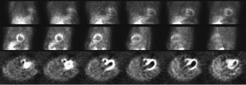 Figure 4.2 Selected projection views (every 15 degrees) from a cardiac SPECT acquisition are shown in the top two rows. In the bottom row are six of the transaxial slices reconstructed from these projections. |
Spatial Resolution
The ideal collimator would have long, small-diameter holes so that only photons arriving exactly perpendicular to the collimator face would be accepted. In reality, the collimator accepts photons over a range of incidence angles. The result is that a small point source would look like a blurred dot in a SPECT image. The amount of blurring depends on the collimator characteristics and the distance of the source from the camera, as depicted in Figure 4.3. The best spatial resolution results when the source is close to the camera and when the collimator has holes that are long or have a small diameter, or both.
Related posts:
 Design Criteria for PET Scanners: What is Important and Why
 11C-Based PET- Radiopharmaceuticals of Clinical Value
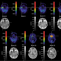 Brain Tumors: Astrocytoma, Oligodendroglioma, and Glioblastoma Images with PET and SPECT
 Sentinel Node Imaging in Head and Neck Tumors
 PET-CT and SPECT-CT of Bone Metastases
 SPECT and PET in Benign Thyroid and Parathyroid Disease
Stay updated, free articles. Join our Telegram channel

Full access? Get Clinical Tree


