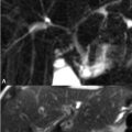
74 Benign Lesions of the Female Pelvis Respiratory and bowel motion are minimal in pelvic imaging, but can degrade image quality if unaccounted for. Fasting should commence 4 to 6 hours prior to pelvic MRI. Administration of intramuscular glucagon may be helpful. Utilization of a compression band to restrict motion may decrease motion-induced artifact, as may respiratory gating, triggering, and utilization of navigator echoes. The bladder should be only moderately distended, in part to minimize truncation artifacts on T2WI. Positive or negative oral contrast is not routinely utilized in MRI in the United States. T2WI but not T1WI illustrate uterine junctional anatomy, distinguishing among the endometrium—high SI due to glandular tissue—junctional zone—hypointense muscular structures—and the myometrium—a moderate SI structure on T2WI during the proliferative phase but hyperintense during the secretory phase due to increased edema and vascular flow. Embryologically, two Müllerian ducts fuse to form the upper vagina, cervix, uterus, and fallopian tubes. Cysts arising from this fusion are similar in MRI appearance and are named by location: nabothian cysts in the cervix; Bartholin gland cysts in the posterolateral vulvovaginal vestibular glands inferior to the pelvic diaphragm; and Gartner cysts, associated with congenital genitourinary abnormalities, above the diaphragm in the anterolateral vagina. Figure 74.1 demonstrates the characteristic appearance of a Bartholin cyst—that is, low SI on (A) axial T1WI and high SI on (B) axial and (C) coronal FS T2WI. Cyst SI varies depending on content, and walls of noninfected cysts do not enhance. Congenital defects in Müllerian duct fusion are reliably characterized on MRI. Class 1 lesions consist of Müllerian hypoplasia or agenesis. The uterus, if present, appears as a low SI structure on T2WI. A unicornuate uterus (class 2) results from unilateral Müllerian duct hypoplasia, the formed uterus appearing as a low volume, off-midline structure. The contralateral horn, if not aplastic, is either cavitary—with approximate preservation of junctional anatomy—or noncavitary— with asymmetric low SI thickening on T2WI. The (A) axial T2WI of Fig. 74.2
![]()
Stay updated, free articles. Join our Telegram channel


Full access? Get Clinical Tree


