
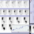
Fig. 21.1
(a–c) Three-phase bone scintigraphy: Phase I (a) showed symmetric flow in the shoulders; in phase II (early phase, b, left image), a mild radiotracer uptake was evident in right shoulder, expression of cellulites; phase III (delayed phase, b, right image) showed moderate radiotracer uptake in right scapulohumeral joint. Whole-body bone scan (c) showed no foci of pathological uptake in other skeletal segments

Fig. 21.2
(a) A normal pattern of radiotracer distribution displayed in pelvis and sacroiliac joint both in early phase (upper image) and in delayed phase (lower image). (b) Scintigraphic pattern of right sacroiliitis: in early phase (upper image), increased blood pool is evident in the right sacroiliac region; in delayed phase (lower image), intense radiotracer uptake is present in right sacroiliac joint, in particular, in its lower part. (c) Scintigraphic pattern of right sacroiliitis, with concomitant arthritis of the hip: in early phase (upper image), increased blood pool is evident in right sacroiliac region; mild radiotracer uptake is present in ipsilateral coxofemoral joint; in delayed phase (lower image), intense and extended radiotracer uptake is evident in right sacroiliac joint, with mild uptake in ipsilateral coxofemoral joint, too


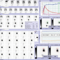
Fig. 21.3
(a–e) Three-phase bone scintigraphy: Phase I (a) showed increased flow in left distal tibia; in phase II (early phase, b, left image), radiotracer uptake in the same site was evident, expression of cellulites; phase III (delayed phase, b, right image) showed intense radiotracer uptake in left tibiotarsal joint, extending to medial malleolus of left tibia; this finding was better defined in lateral (d) and anterior static views of the feet (e). Whole-body bone scan showed no foci of pathological uptake in other skeletal segments (c)

Fig. 21.4
(a–c) Early phase whole-body scan. (a) Increased radiotracer uptake in left knee was evident, indicating cellulitis; delayed phase whole-body scan (b) showed radiotracer uptake in the same site, extending to the region above the metaphysis in ipsilateral distal femur, indicating arthritis complicated by osteomyelitis. This finding was better defined in static views of the knees (c). Delayed whole-body scan (b) showed no foci of pathological uptake in other skeletal segments


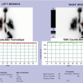
Fig. 21.5


(a–c) Three-phase bone scintigraphy: in phase II (early phase, a, images on the left), severe and extended reduction of radiotracer uptake was evident in left hip joint, as in extended edema; phase III (delayed phase, b, images on the right) showed severe reduction of radiotracer uptake in head and neck of left femur, because of severe compressive surrounding edema. Whole-body bone scan (c) showed mild increased radiotracer uptake in the region above the metaphysis in ipsilateral distal femur, indicating another focus of osteomyelitis
Related posts:


Stay updated, free articles. Join our Telegram channel
Full access? Get Clinical Tree
