Fig. 1.1
Pulsion diverticulum in a 66-year-old female. (a) Esophagography shows middle esophageal diverticulum with a wide neck and smooth contour, which are typical findings of pulsion diverticulum. (b) As pulsion diverticulum does not have muscle layer in the wall, barium usually remains at the diverticulum after the esophagus has emptied
1.8.2 Killian-Jamieson
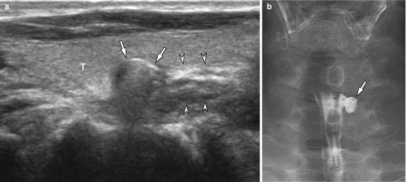
Fig. 1.2
The Killian-Jamieson diverticulum in a 72-year-old female. (a) Ultrasonography longitudinally scanning the left thyroid gland (T) shows a round hypoechoic nodule with an arc-shape hyperechoic portion with posterior reverberation (arrows). Note the contiguity with the esophagus (arrowheads). (b) Esophagography shows a well-circumscribed outpouching structure (arrow) protruding from the left lateral wall of the cervical esophagus
1.8.3 Epiphrenic Diverticulum
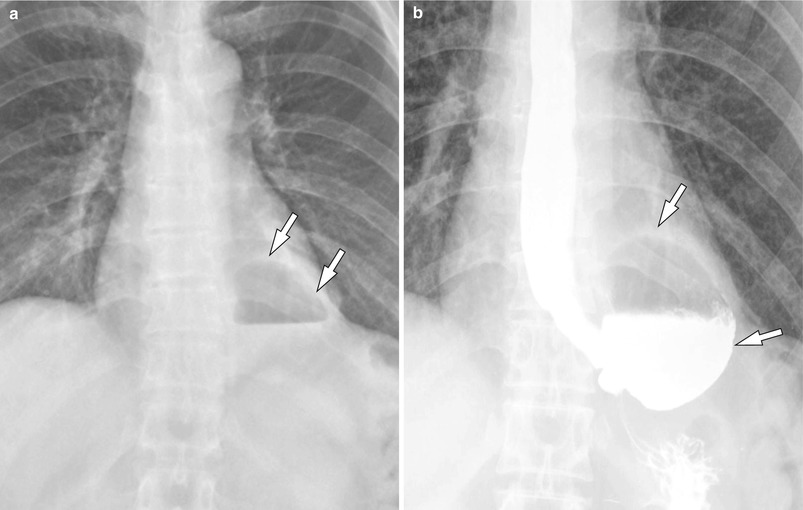
Fig. 1.3
Epiphrenic diverticulum in a 52-year-old female. (a) Posteroanterior chest radiography shows a cavitary lesion with air-fluid level (arrows) at the retrocardiac space. (b) A barium esophagography reveals a large diverticulum (arrows) arises in the distal esophagus
1.8.4 Hiatal Hernia (Type I; Sliding Hiatal Hernia)
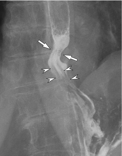
Fig. 1.4
Hiatal hernia (type I, sliding hiatal hernia) in an 86-year-old female. Note the upward displacement of the gastroesophageal junction (arrows) and gastric rugae (arrowheads)
1.8.5 Hiatal Hernia (Type III)
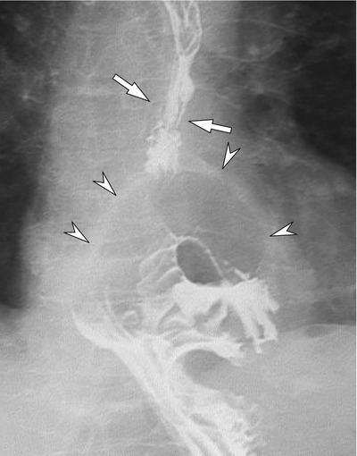
Fig. 1.5
Hiatal hernia (type III) in an 86-year-old female. With upward displacement of the gastroesophageal junction (arrows), herniation of the gastric fundus (arrowheads) is also seen on esophagography
1.8.6 Uphill Esophageal Varix
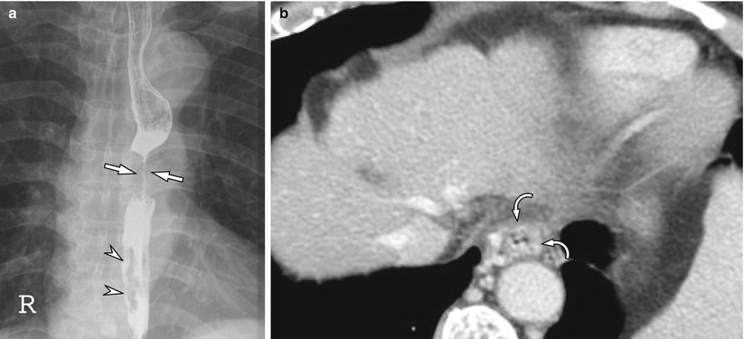
Fig. 1.6
Uphill esophageal varix in a 68-year-old male with esophageal carcinoma. (a) With circumferentially infiltrating carcinoma of the middle esophagus (arrows), the varices are seen as serpiginous filling defects in the distal esophagus (arrowheads). (b) Contrast-enhanced transverse CT image shows a cirrhotic liver and prominent varix (curved arrows) in the distal esophagus
1.8.7 Downhill Esophageal Varix
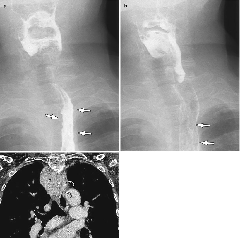
Fig. 1.7


Downhill esophageal varix in a 76-year-old female. (a, b) Note the left-side displacement of the proximal esophagus and thickened, nodular folds (arrows). (c) Contrast-enhanced coronal CT image shows a large intrathoracic goiter (G), which causes the mass effect to the adjacent organs. Esophageal varices localized to the proximal esophagus (curved arrows) are also seen as serpentine structures with homogeneous enhancement
Related posts:
Stay updated, free articles. Join our Telegram channel
Full access? Get Clinical Tree
