Fig. 10.1
Intestinal malrotation (nonrotation type) in a 39-year-old man. (a, b) The jejunum is directly continued from the duodenum (arrow in a), with absence of the normal 3rd and 4th segment of the duodenum and duodenojejunal junction (asterisks in b). (c, d) The small bowel loops (straight arrows) lie in the right side of the abdomen, and the colon (curved arrows) lies in the left side
10.4.2 Midgut Volvulus
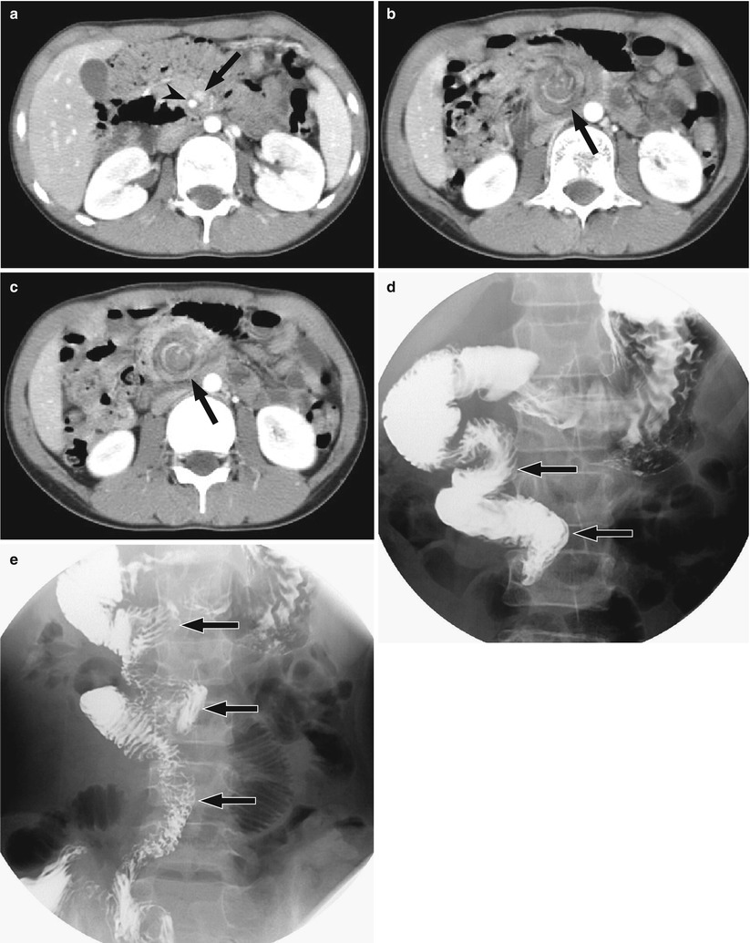
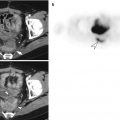
Fig. 10.2
Midgut volvulus in an 11-year-old boy. (a) Axial contrast-enhanced CT shows transposition of superior mesenteric artery (arrowhead) and vein (arrow). (b, c) Axial CT below (a) shows “whirlpool sign” (arrow) which consists of wrapping of the superior mesenteric vein and its branches, mesenteric fat, bowel loops around the superior mesenteric artery in the clockwise direction. (d, e) UGIS shows “corkscrew sign” (arrows), a spiral configuration of the 4th portion of the duodenum and proximal jejunum
10.4.3 Meckel Diverticulitis
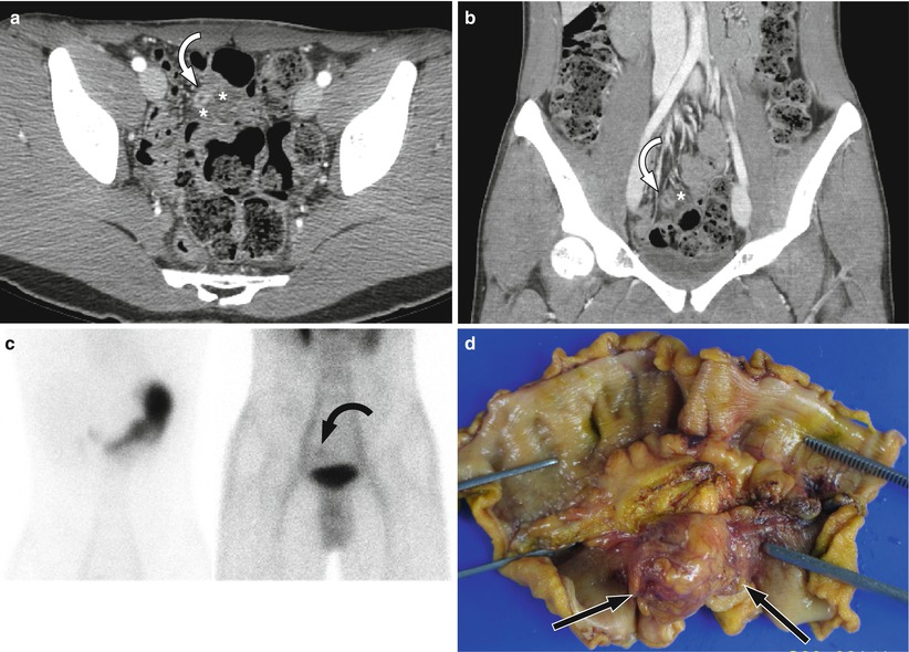
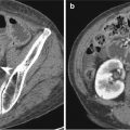
Fig. 10.3
Meckel diverticulitis in a 23-year-old man. (a, b) Axial (a) and coronal (b) contrast-enhanced CT shows a tubular structure (curved arrow) with enhancing thick wall adjacent to distal small bowel loops. Note fat stranding (asterisks) around the tubular-enhancing structure. (c) Tc-99m pertechnetate scintigraphy shows a persistent focus of uptake (curved arrow) in the right upper side of the urinary bladder, suggesting the presence of heterotopic gastric mucosa. (d) Gross specimen shows inflamed Meckel diverticulum (arrows)
10.4.4 Meckel Diverticulum
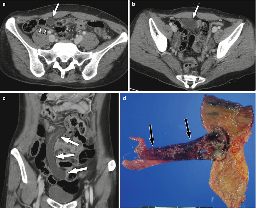
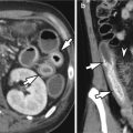
Fig. 10.4
Meckel diverticulum in a 51-year-old woman. (a, b) Axial contrast-enhanced CT shows a fluid-filled tubular structure coursing in a craniocaudal direction. Note normal appendix (arrowheads). (c) Coronal CT shows a large fluid-filled curved tubular structure (arrows) in the abdomen. (d) Gross specimen shows a giant Meckel diverticulum (arrows) arising from the ileum
10.4.5 Meckel Diverticulum with Small Bowel Obstruction
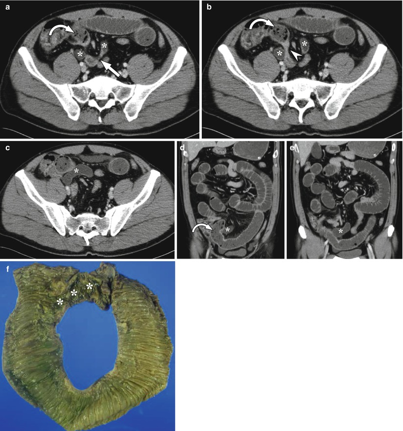
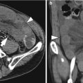
Fig. 10.5


Meckel diverticulum with small bowel obstruction in a 48-year-old man. (a–e) Axial and coronal contrast-enhanced CT images show a fluid-filled curved tubular structure (asterisks) which originates from the distal ileum (arrow). Diffuse fluid-filled small bowel dilation is seen, which suggested small bowel obstruction. The transitional point (arrowhead) is noted near the origin site of the tubular structure, with the proximal small bowel feces sign (curved arrow). (f) Gross specimen of the resected small bowel shows a Meckel diverticulum (asterisks) originating from the ileum
Related posts:

Stay updated, free articles. Join our Telegram channel
Full access? Get Clinical Tree
