Fig. 4.1
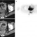
Gastric duplication cyst in a 28-year-old male. (a) On CT, an approximately 5 cm subepithelial cystic mass (white arrow) is seen at the posterior wall of the gastric fundus. (b) On EUS, this mass (asterisk) manifests anechoic cystic nature and the presence of an echogenic inner rim with hypoechoic outer muscle layers (“muscular rim” sign). (c) Endoscopy demonstrates a large subepithelial tumor in the gastric fundus. (d) Pathologic examination reveals this mass as the foregut duplication cyst
4.7.2 Duodenal Duplication Cyst
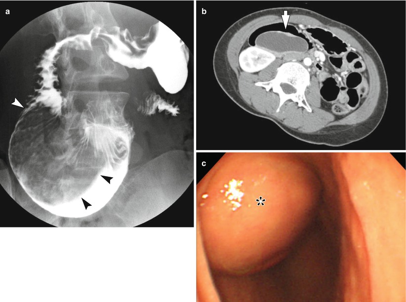
Fig. 4.2
Duodenal duplication cyst in a 22-year-old female. (a) A huge filling defect is noted in the dilated duodenal second and third portions (arrowheads) in UGIS. (b) On CT, a large and well-defined cystic mass is located in the medial wall of the duodenal second portion (arrow). (c) Endoscopy shows a well-defined subepithelial tumor (asterisk) in the duodenal second portion with intact overlying mucosa
4.7.3 Gastric Diverticulum
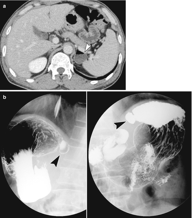
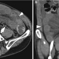
Fig. 4.3
Gastric diverticulum. (a) A case of gastric diverticulum (arrowhead) in a 56-year-old male. If there was no air bubble within the diverticulum, it could mimic the left adrenal gland mass or gastric subepithelial tumor on CT. (b) Gastric diverticula (arrowheads) on UGIS. A diagnosis of gastric diverticulum can be made if pooling of barium in the diverticulum with typical location of posterior wall of the fundus is observed
4.7.4 Duodenal Diverticulum
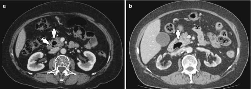
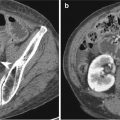
Fig. 4.4
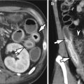
Duodenal diverticulum. (a) Two small duodenal diverticula (arrows) are located on the medial border of the second portion of duodenum, the periampullary region. When duodenal diverticulum contains fluid predominantly, it can mimic a cystic neoplasm in the pancreatic head. (b) An approximately 3 cm duodenal diverticulum with internal bubble lucency is located on the medial wall of the duodenal second portion (arrow)
4.7.5 Diagram of the Two Main Types of Gastric Volvulus
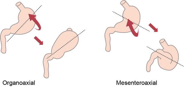
Fig. 4.5
Gastric volvulus. Diagram illustrates the two main types of gastric volvulus (organoaxial and mesenteroaxial)
4.7.6 Organoaxial Gastric Volvulus
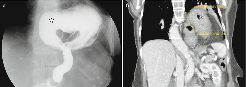
Fig. 4.6


Organoaxial gastric volvulus in a 94-year-old male. (a) The gastric antrum (asterisk) moves from an inferior to a superior position on UGIS with hiatal hernia. (b) The coronal CT scan shows the same findings
Related posts:

Stay updated, free articles. Join our Telegram channel
Full access? Get Clinical Tree
