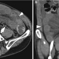
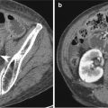
Fig. 14.1
Ileal hemangioma in an 18-year-old male. (a) Contrast-enhanced transverse CT image shows a soft tissue mass (arrows) with contrast enhancement in the central area. (b) Surgical specimen of segmental ileal resection shows a round mass (arrows)
14.10.2 Ileal Leiomyoma
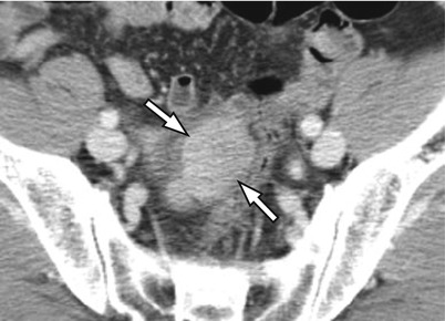
Fig. 14.2
Ileal leiomyoma in a 60-year-old male. Contrast-enhanced transverse CT image shows a soft tissue mass (arrows) with moderate contrast enhancement
14.10.3 Ileal Lipoma
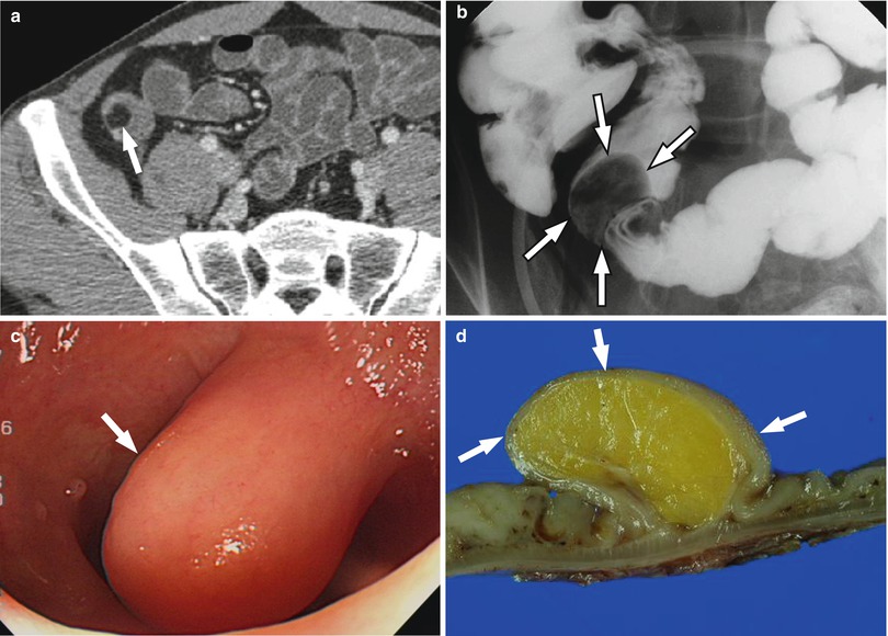
Fig. 14.3
Ileal lipoma in an 18-year-old male. (a) Contrast-enhanced transverse CT image shows a fat-attenuating intraluminal mass (arrow) in the small bowel. (b) Colon study with ileal reflux shows a filling defect in the ileum (arrows). (c) Colonoscopy with ileal intubation shows an intraluminal round mass (arrow). (d) Surgical specimen of segmental ileal resection shows a round mass (arrows) with internal fat
14.10.4 Peutz-Jeghers Syndrome with Duodenal Cancer with Hepatic Metastases
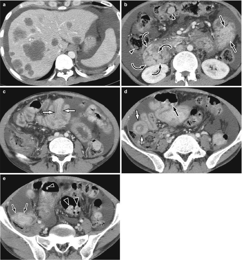
Fig. 14.4


A 34-year-old male with Peutz-Jeghers syndrome with duodenal cancer with hepatic metastases. Contrast-enhanced transverse CT images show the following findings. (a) Multiple hepatic nodules representing disseminated metastases. (b) Duodenal mass with irregular luminal surface representing duodenal cancer (curved arrows), large polypoid mass in the jejunum (dark arrows), and multiple colon polyps (arrowheads). (c) Small bowel intussusception (white arrows) associated with polyp (not seen). (d) Small bowel intussusception (white arrows) and small bowel polyp (dark arrow). (e) Polyps in the small bowel (dark arrows) and colon (arrowheads)
Stay updated, free articles. Join our Telegram channel

Full access? Get Clinical Tree


